Copyright
©The Author(s) 2016.
World J Gastroenterol. May 21, 2016; 22(19): 4626-4637
Published online May 21, 2016. doi: 10.3748/wjg.v22.i19.4626
Published online May 21, 2016. doi: 10.3748/wjg.v22.i19.4626
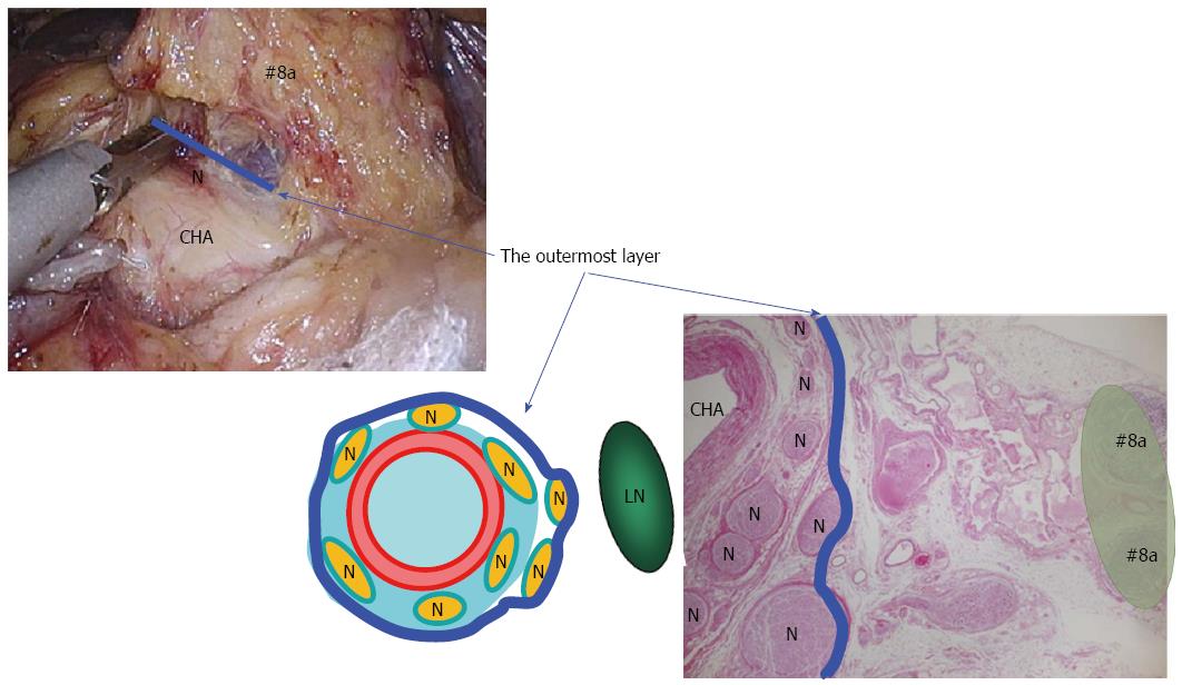
Figure 1 Outermost layer of the autonomic nerve.
Shown in the blue line, lies between the vascular sheath of the major arteries and the fat tissue including lymph nodes. Appropriate tension given to this thin loose connective tissue layer generates sufficient space for safe, adequate and reproducible prophylactic lymph node dissection along the major arteries. LN: Lymph node; N: Nerve; CHA: Common hepatic artery.
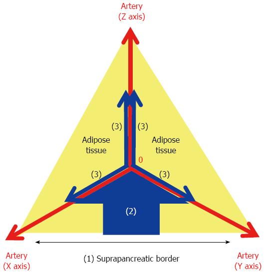
Figure 2 XYZ-axis theory.
The following three steps result in effective probing of the outermost layer: (1) dissection of the serosal membrane on the suprapancreatic border; (2) dissection of the fat tissue in the caudo-cranial direction towards the zero point; and (3) dissection of the fat tissue bearing the target LNs in the medio-lateral direction along the outermost layer on the XZ and YZ axes. The outermost layer adjacent to the zero point should be exposed during the 2nd step.
Figure 3 Lymph node dissection along the outermost layer using the XYZ-axis theory.
A: No. 7 and 9 dissection, B: No. 6 dissection, C: No. 5 dissection.
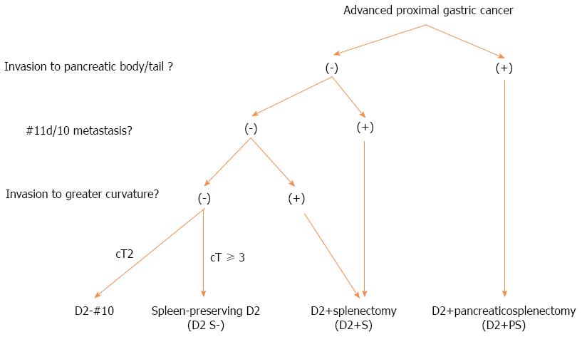
Figure 4 Indication for splenic hilar lymph node dissection at FHU.
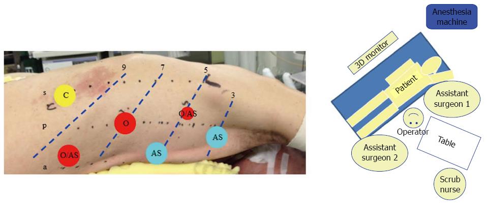
Figure 5 Setup for prone VATS-E at Fujita Health University.
A: The patient in the hemi-prone position using the six-trocar system. 12 mm trocars are used except for the trocar in the 5th intercostal space (ICS) behind the posterior axillary line; B: OR setup. s: Scapula angle line; p: Posterior axillary line; a: Anterior axillary line; 3: 3rd ICS; 5: 5th ICS; 7: 7th ICS; 9: 9th ICS; O: Operating surgeon; AS: Assistant surgeon.
- Citation: Suda K, Nakauchi M, Inaba K, Ishida Y, Uyama I. Minimally invasive surgery for upper gastrointestinal cancer: Our experience and review of the literature. World J Gastroenterol 2016; 22(19): 4626-4637
- URL: https://www.wjgnet.com/1007-9327/full/v22/i19/4626.htm
- DOI: https://dx.doi.org/10.3748/wjg.v22.i19.4626


