Copyright
©The Author(s) 2016.
World J Gastroenterol. May 21, 2016; 22(19): 4619-4625
Published online May 21, 2016. doi: 10.3748/wjg.v22.i19.4619
Published online May 21, 2016. doi: 10.3748/wjg.v22.i19.4619
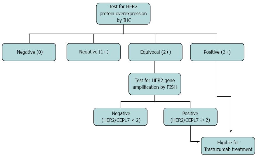
Figure 1 Human epidermal growth factor receptor 2 testing algorithm.
HER2: Human epidermal growth factor receptor 2; IHC: Immunohistochemistry; FISH: Fluorescent in situ hybridization; CEP17: Chromosome 17.
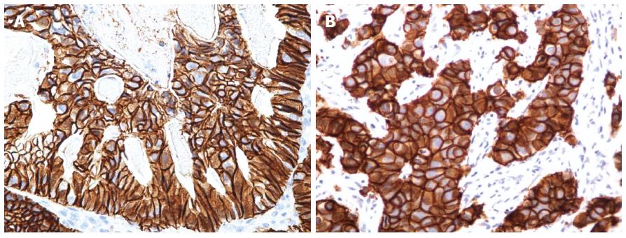
Figure 2 Human epidermal growth factor receptor 2 expression in gastric and breast tumors.
A: A HER2-positive (3+) case of gastric adenocarcinoma; the cytoplasmic membranous immunostaining is incomplete and predominantly basolateral (× 400); B: A HER2-positive (3+) case of invasive ductal carcinoma of the breast; the cytoplasmic membranous staining is fully circumferential (× 400). HER2: Human epidermal growth factor receptor 2.
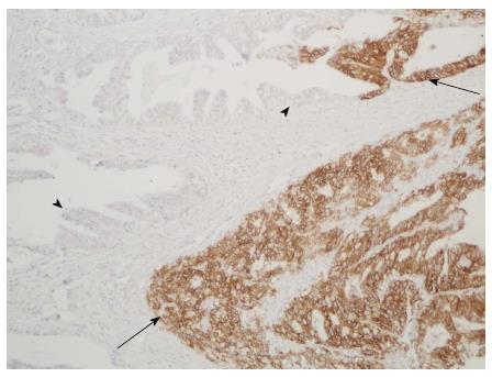
Figure 3 Representative image of the intratumoral heterogeneity of HER2 expression.
Arrows indicate areas with strong continuous membranous staining (score 3+) and arrowheads indicate negative areas (score 0) (× 100). HER2: Human epidermal growth factor receptor 2.
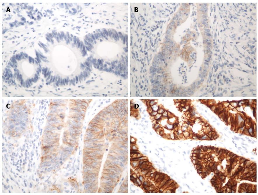
Figure 4 Human epidermal growth factor receptor 2 protein expression in gastric and gastroesophageal tumors.
A: A negative (0) case; B: A negative (+1) case; C: An equivocal (2+) case; D: A positive (3+) case. HER2: Human epidermal growth factor receptor 2.
- Citation: Abrahao-Machado LF, Scapulatempo-Neto C. HER2 testing in gastric cancer: An update. World J Gastroenterol 2016; 22(19): 4619-4625
- URL: https://www.wjgnet.com/1007-9327/full/v22/i19/4619.htm
- DOI: https://dx.doi.org/10.3748/wjg.v22.i19.4619


