Copyright
©The Author(s) 2016.
World J Gastroenterol. May 14, 2016; 22(18): 4604-4609
Published online May 14, 2016. doi: 10.3748/wjg.v22.i18.4604
Published online May 14, 2016. doi: 10.3748/wjg.v22.i18.4604
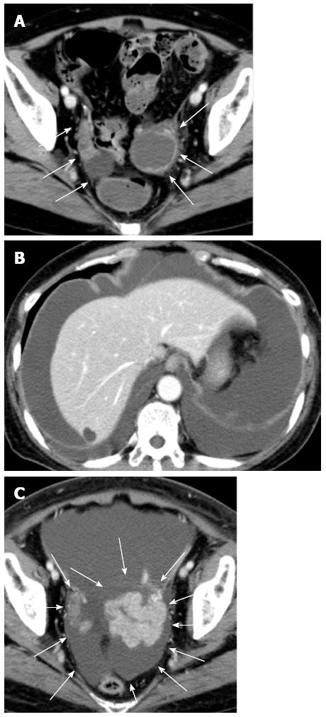
Figure 1 Computed tomography of the abdomen.
A: The examination performed 22 d before admission showed bilateral enlarged ovaries with solid and cystic components (arrows). Ascites was not visible; B and C: The examination performed 26 d after admission revealed massive ascites and pleural effusion and rapid enlargement of ovarian tumors (arrows).
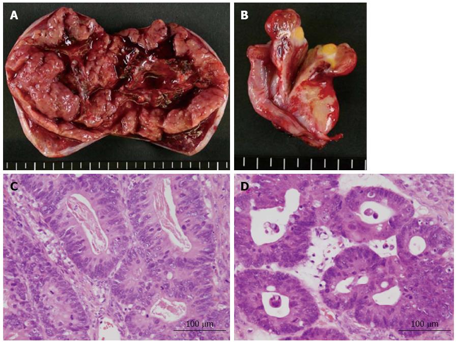
Figure 2 Pathological analysis.
A, B: Cross-sectional views of the left and right ovarian tumors, respectively. The left ovarian tumor measured 90 mm × 55 mm, and the right ovarian tumor measured 30 mm × 30 mm. Both tumors contained solid and cystic components; C, D: Microscopic views of the left and right ovarian tumors, respectively, showing moderately differentiated adenocarcinoma.
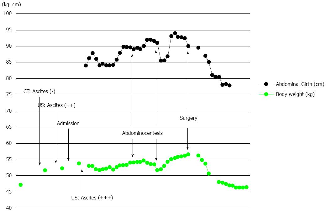
Figure 3 Line graphs showing changes in body weight and abdominal girth.
Note that the body weight increased more than 4 kg before the emergence of ascites and that both the body weight and abdominal girth did not change for 3 d after surgery but thereafter decreased rapidly. CT: Computed tomography; US: Ultrasound.
- Citation: Kyo K, Maema A, Shirakawa M, Nakamura T, Koda K, Yokoyama H. Pseudo-Meigs’ syndrome secondary to metachronous ovarian metastases from transverse colon cancer. World J Gastroenterol 2016; 22(18): 4604-4609
- URL: https://www.wjgnet.com/1007-9327/full/v22/i18/4604.htm
- DOI: https://dx.doi.org/10.3748/wjg.v22.i18.4604


