Copyright
©The Author(s) 2016.
World J Gastroenterol. May 14, 2016; 22(18): 4567-4575
Published online May 14, 2016. doi: 10.3748/wjg.v22.i18.4567
Published online May 14, 2016. doi: 10.3748/wjg.v22.i18.4567
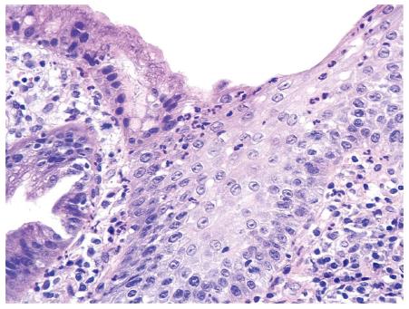
Figure 1 Islets of non-keratinized squamous epithelium can be observed intermingling with cardiac-type gastric mucosa.
Superficial and glandular gastric epithelial linings demonstrate infiltration by polymorphonuclear leukocytes (Hematoxylin-eosin staining; original magnification × 10).
Figure 2 Histological section of gastric-type columnar mucosa of the esophagus.
Moderate mononuclear infiltrate in the lamina propria and polymorphonuclear leukocytes infiltrating the mucosa layer can be observed (Hematoxylin-eosin staining stain; original magnification × 40).
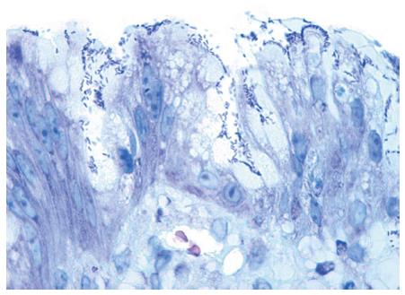
Figure 3 Numerous rod-shape bacilli can be seen attached to the gastric-type metaplastic columnar epithelium (Giemsa staining; original magnification × 100).
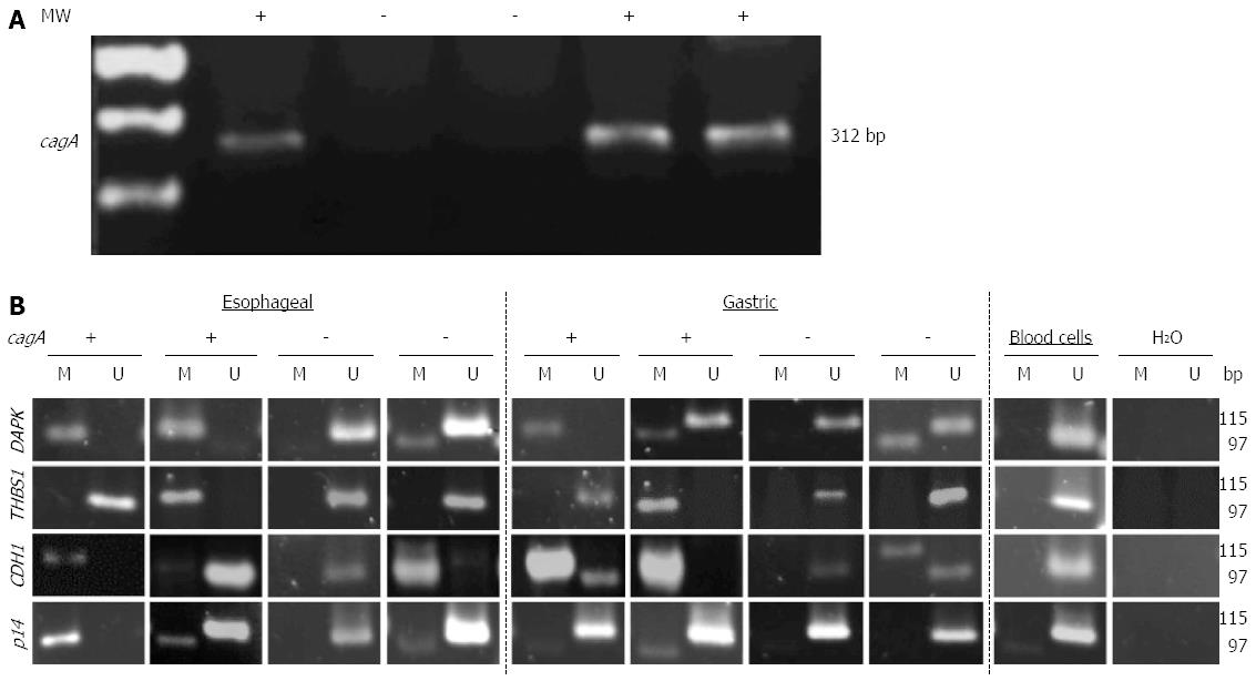
Figure 4 cagA detection and DNA promoter methylation status of DAPK, THBS1, CDH1 and p14 genes in esophageal and gastric mucosa tissue samples.
A: Helicobacter pylori cagA+ detection by polymerase chain reaction (PCR) amplification in tissue samples, (+) positive and (-) negative; B: Analysis of methylation status of DAPK, THBS1, CDH1, and p14 genes promoters in esophageal mucosa and gastric biopsies tissue samples detected by methyl-sensitive PCR. Blood cells were used as control of unmethylated promoter regions. M represents methylated and U represent unmethylated; the status of Helicobacter pylori cagA is indicated as (+) positive and (-) negative.
- Citation: Herrera-Goepfert R, Oñate-Ocaña LF, Mosqueda-Vargas JL, Herrera LA, Castro C, Mendoza J, González-Barrios R. Methylation of DAPK and THBS1 genes in esophageal gastric-type columnar metaplasia. World J Gastroenterol 2016; 22(18): 4567-4575
- URL: https://www.wjgnet.com/1007-9327/full/v22/i18/4567.htm
- DOI: https://dx.doi.org/10.3748/wjg.v22.i18.4567


