Copyright
©The Author(s) 2016.
World J Gastroenterol. May 14, 2016; 22(18): 4427-4437
Published online May 14, 2016. doi: 10.3748/wjg.v22.i18.4427
Published online May 14, 2016. doi: 10.3748/wjg.v22.i18.4427
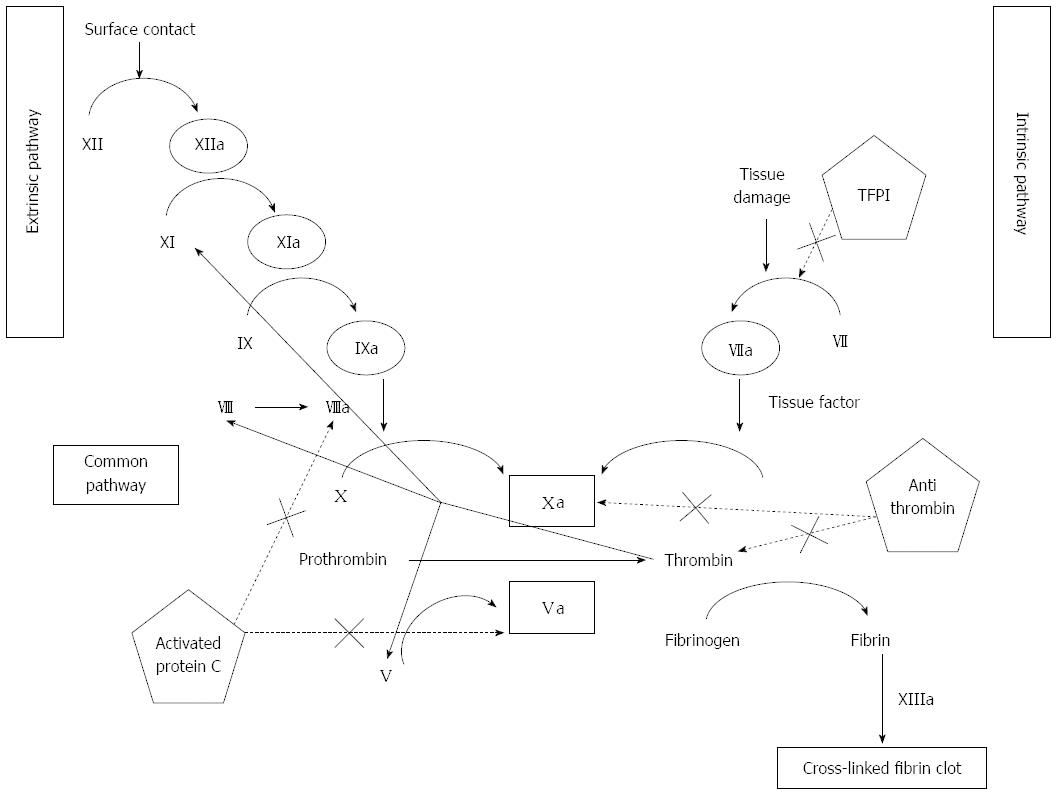
Figure 1 Coagulation cascade: Extrinsic, intrinsic, and common pathways.
Coagulation factors are depicted in roman numerals with the suffix “a” denoting activated forms of the factors. Inhibitory pathways are shown in dotted lines. TFPI: Tissue factor pathway inhibitor.
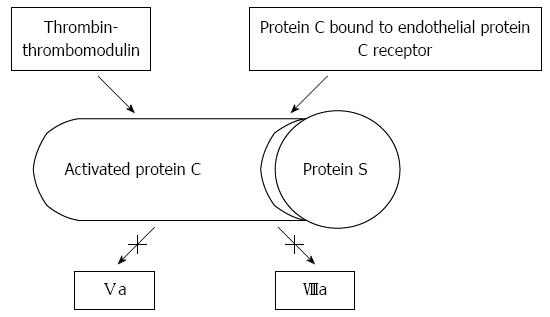
Figure 2 Anticoagulant effect of thrombomodulin, protein C, and protein S.
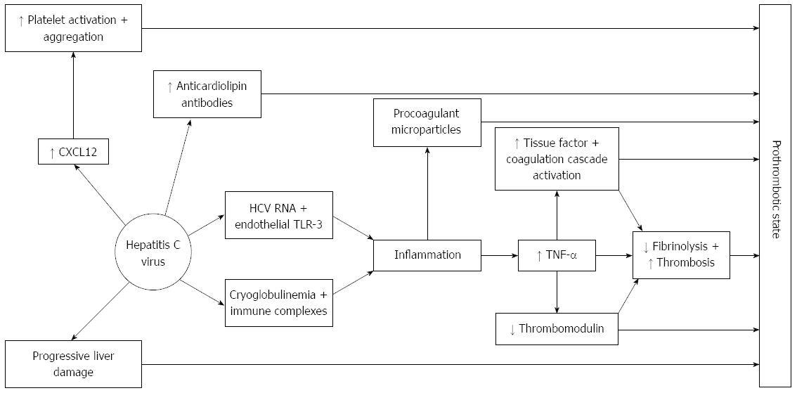
Figure 3 Hepatitis C virus has several effects on anticoagulant and procoagulant cascades, in addition to the effects on coagulation derived from liver cirrhosis development.
(1) Hepatitis C virus (HCV) infection is associated with anticardiolipin antibodies which are related to thrombotic events; (2) Viral HCV RNA binds to toll-like receptors (TLR)-3 found in endothelial cells which leads to inflammation. Also, HCV infection is associated with cryoglobulinemia and thus immune complexes directed against viral RNA are formed. Inflammation generated by these two mechanisms lead to TNF-α secretion. TNF-α is an inducer of tissue factor expression, therefore exerting a prothrombotic effect by activating the coagulation cascade and also downregulates thrombomodulin expression; and (3) In HCV infection, CXCL12 is up-regulated in the endothelium of blood vessels formed in active inflammatory foci. CXCL12 is a potent promoter of platelet aggregation and adhesion.
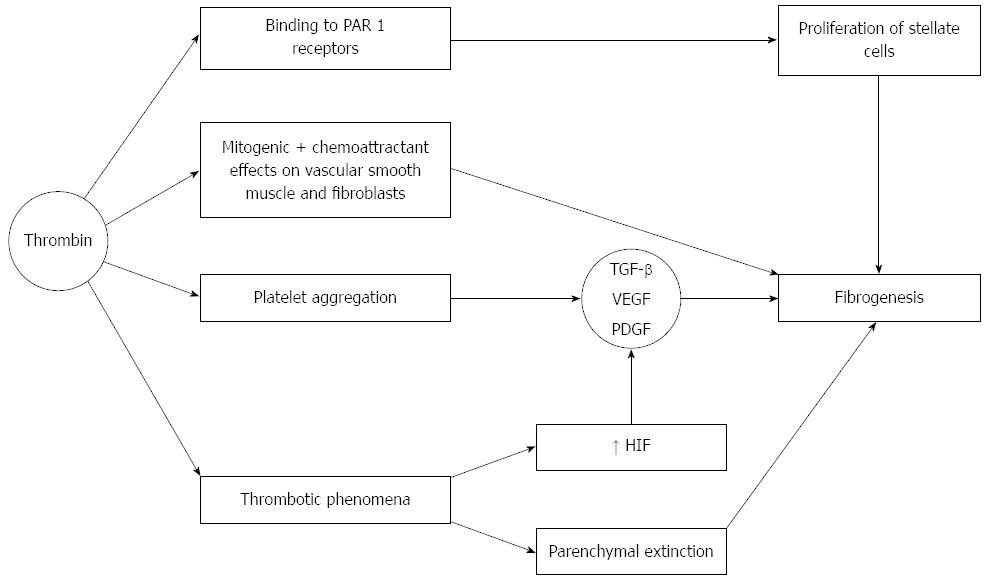
Figure 4 Effects on thrombin on fibrogenesis.
Thrombin binds to PAR-1 receptors on hepatic stellate cells which leads to proliferaton and activation of these cells Thrombin also promotes platelet aggregation. Platelet alpha granules are rich in several growth factors, including TGF-β, which in turn promotes fibrogenesis. Vascular endothelial growth factor (VEGF) and PDGF also play contributory roles. Thrombin also exerts mitogenic and chemoattractant effects on vascular smooth muscle cells and fibroblasts. Finally, thrombotic phenomena also occur within the liver, leading to ischemic parenchymal injury, and substitution of parenchyma by fibrous tissue (the so called parenchymal extinction). When a clot provokes ischemia, VEGF, PDGF and TGF-β are activated probably via an increase in hypoxia-inducible-factor (HIF), which is raised in cirrhosis in relation to portal microthrombotic phenomena.
- Citation: González-Reimers E, Quintero-Platt G, Martín-González C, Pérez-Hernández O, Romero-Acevedo L, Santolaria-Fernández F. Thrombin activation and liver inflammation in advanced hepatitis C virus infection. World J Gastroenterol 2016; 22(18): 4427-4437
- URL: https://www.wjgnet.com/1007-9327/full/v22/i18/4427.htm
- DOI: https://dx.doi.org/10.3748/wjg.v22.i18.4427


