Copyright
©The Author(s) 2016.
World J Gastroenterol. Apr 21, 2016; 22(15): 4020-4026
Published online Apr 21, 2016. doi: 10.3748/wjg.v22.i15.4020
Published online Apr 21, 2016. doi: 10.3748/wjg.v22.i15.4020
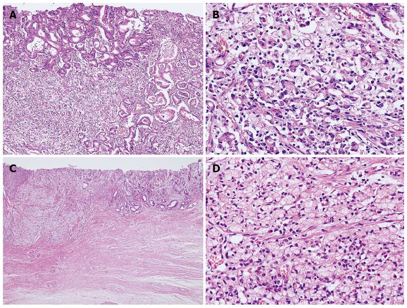
Figure 1 Pathology of mixed-type gastric carcinoma.
A: Mixed-type gastric carcinoma showing moderately differentiated tubular adenocarcinoma, intestinal type (upper left and right) and poorly differentiated adenocarcinoma (lower left); B: High magnification view of poorly differentiated lesion (× 400); C: Mixed-type gastric carcinoma showing moderately differentiated tubular adenocarcinoma, intestinal type (right) and signet ring cell carcinoma (left); D: High magnification view of signet ring cell carcinoma lesion (× 400).
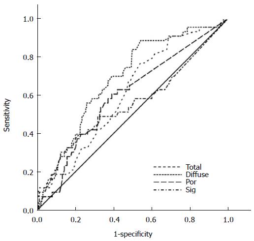
Figure 2 Receiver operating characteristics curve analysis of scores for predicting lymph node metastasis in mixed-type early gastric cancers yielded an area under the curve of 0.
687 (95%CI: 0.604-0.701, P < 0.0001) for the diffuse component, 0.610 (95%CI: 0.523-0.698, P = 0.023) for total size, 0.598 (95%CI: 0.505-0.691, P = 0.043) for the poorly differentiated component, and 0.556 (95%CI: 0.450-0.661, P = 0.249) for the signet ring cell component.
- Citation: Hwang CS, Ahn S, Lee BE, Lee SJ, Kim A, Choi CI, Kim DH, Jeon TY, Kim GH, Song GA, Park DY. Risk of lymph node metastasis in mixed-type early gastric cancer determined by the extent of the poorly differentiated component. World J Gastroenterol 2016; 22(15): 4020-4026
- URL: https://www.wjgnet.com/1007-9327/full/v22/i15/4020.htm
- DOI: https://dx.doi.org/10.3748/wjg.v22.i15.4020


