Copyright
©The Author(s) 2016.
World J Gastroenterol. Apr 21, 2016; 22(15): 3992-4001
Published online Apr 21, 2016. doi: 10.3748/wjg.v22.i15.3992
Published online Apr 21, 2016. doi: 10.3748/wjg.v22.i15.3992
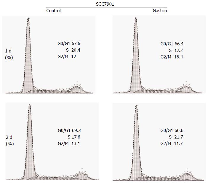
Figure 1 Gastrin-stimulated translocation of calcyclin binding protein/Siah-1 interacting protein into nucleus decreases the number of SGC7901 gastric cancer cell in the G0-G1 phases of the cell cycle.
Cells were treated with gastrin (10-8 mol/L) for the indicated times and cell cycle variables were investigated by flow cytometry after propidium iodide (PI) staining. Data are presented as mean ± SD (n = 3), and graphs shown are representative of the three experiments.
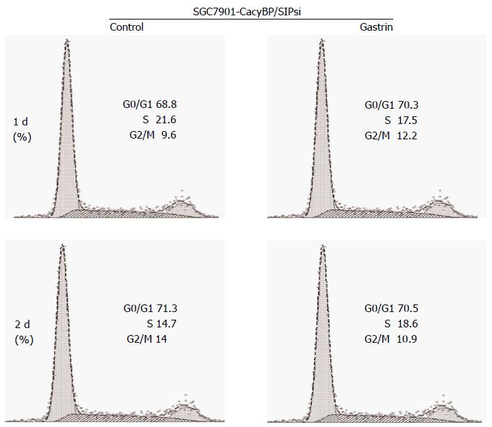
Figure 2 Treatment with gastrin increases the number of SGC7901-calcyclin binding protein/Siah-1si1 cells in the G0-G1 phases of the cell cycle.
Cells were treated with gastrin (10-8 mol/L) for the indicated times and cell cycle variables were investigated by flow cytometry. Data are presented as mean ± SD (n = 3), and graphs shown are representative of the three experiments.
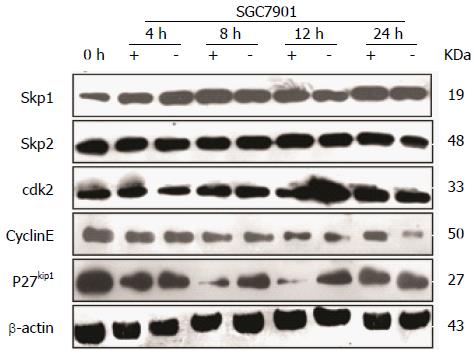
Figure 3 Effects of calcyclin binding protein/Siah-1 on cell cycle regulatory proteins.
Cells were synchronized in G2-M phase with 0.2 μg/mL nocodazole for 15 h and nocodazole was removed by washing; cells were then incubated in fresh medium with (+) or without (-) gastrin for the indicated times. After treatment, cellular lysates were prepared and loaded per lane. Different blots with the same samples were detected with the indicated antibodies: Cyclin E, CDK2, p27Kip1, Skp1, Skp2, and β-actin as an internal control. Gastrin treatment induced an increase in the amount of Cyclin E protein and a decrease in the level of p27Kip1 protein during the first 8 h of treatment, whereas the levels of Skp1, Skp2, and CDK2 were not affected.
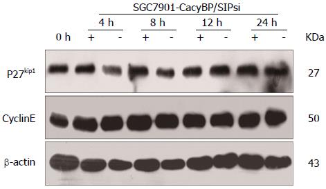
Figure 4 No change in protein levels of p27Kip1 and Cyclin E was observed in SGC7901-calcyclin binding protein/Siah-1si1 cells in which expression of calcyclin binding protein/Siah-1 was silenced.
Cells were synchronized in G2-M phase with nocodazole for 15 h and nocodazole was removed by washing; and then cells were incubated in fresh medium with (+) or without (-) gastrin (10-8 mol/L). Different blots with the same samples were detected with the indicated antibodies: Cyclin E, p27Kip1, and β-actin as an internal control.
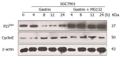
Figure 5 Proteasome-mediated degradation of p27Kip1 in gastric cancer cells.
Western blot analysis of p27Kip1, Cyclin E and β-actin in cells 4, 8, 12, and 24 h after treatment with 10-8 mol/L gastrin in the absence or presence of MG132 (5 mmol/L, 15 min pre-treatment and co-treatment with gastrin).
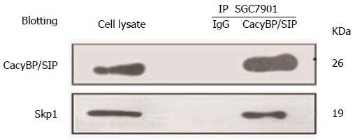
Figure 6 Physical interactions between Skp1 and calcyclin binding protein/Siah-1 in SGC7901 cells.
Nuclear protein extracts from gastric cancer cells treated with 10-8 mol/L gastrin were immunoprecipitated with calcyclin binding protein/Siah-1 (CacyBP/SIP) MAb or control IgG. Immune complexes were analyzed by immunoblotting using an anti-Skp1 antibody with ECL-based detection.
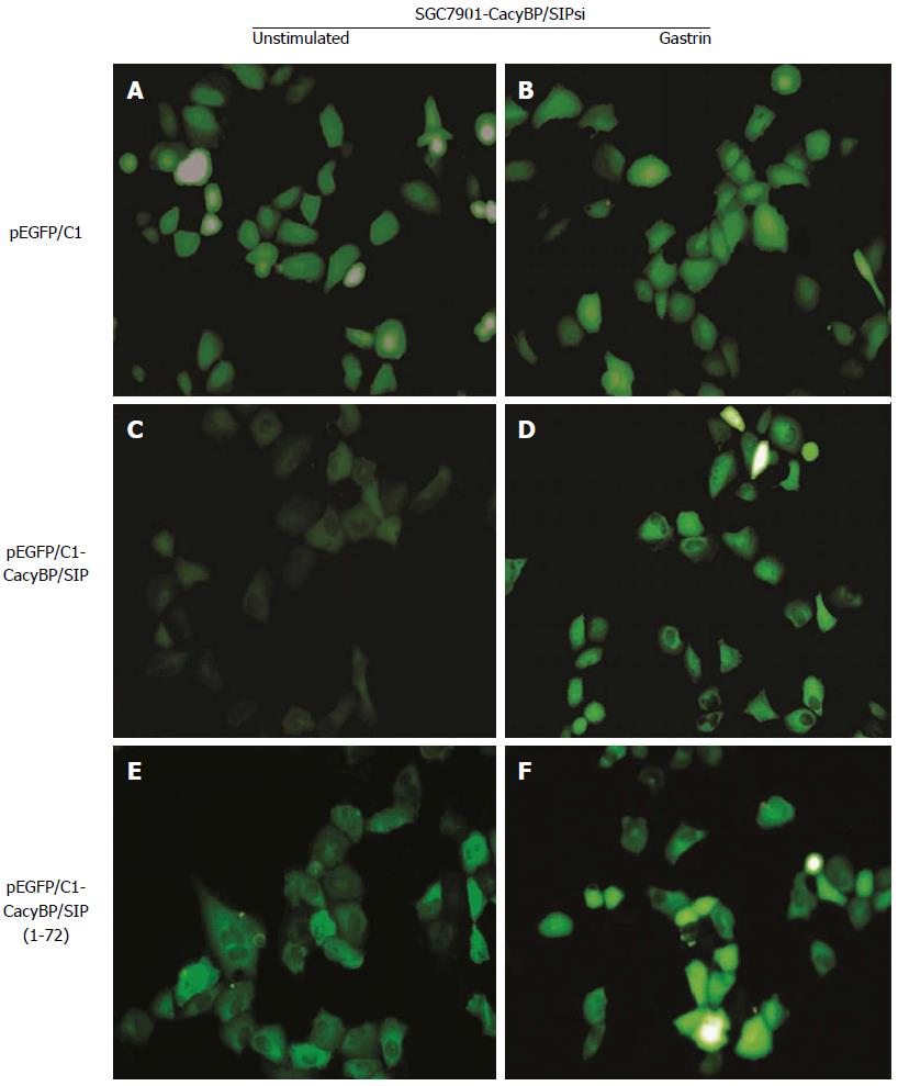
Figure 7 Immunofluorescent localization of calcyclin binding protein/Siah-1 (1-72) in SGC7901-calcyclin binding protein/Siah-1si cells.
SGC7901-CacyBP/SIPsi cells were transfected with pEGFP/C1 vector, producing either GFP-tagged CacyBP/SIP or CacyBP/SIP (1-72) lacking the 155 central and C-terminal amino acids. Cells transfected with GFP-tagged pEGFP/C1 lacking a cDNA insert served as controls. Transfectants were treated or untreated with gastrin (10-8 mol/L), and EGFP fluorescence was analyzed under a confocal laser microscope. A, C, E: Unstimulated cells; B, D, F: Cells 8 h after gastrin stimulation.
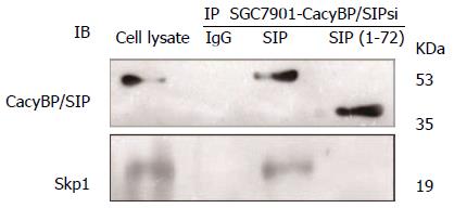
Figure 8 Interaction between Skp1 and calcyclin binding protein/Siah-1 (1-72) in SGC7901-calcyclin binding protein/Siah-1si transfected with pEGFP/C1-calcyclin binding protein/Siah-1 (1-72).
Nuclear protein extracts from cells transfected with pEGFP/C1-CacyBP/SIP or pEGFP/C1- CacyBP/SIP (1-72) after treatment with 10-8 mol/L gastrin were immunoprecipitated with anti-GFP antibody or control IgG. Immune complexes were analyzed by immunoblotting using an anti-Skp1/GFP antibody with ECL-based detection. CacyBP/SIP: Calcyclin binding protein/Siah-1 interacting protein.
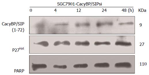
Figure 9 Lysates of the transfected cells were harvested for Western blot analysis 0, 4, 12, 24, and 48 h after treatment with gastrin (10-8 mol/L).
Equal amounts of cellular protein (40 μg) were subjected to SDS-PAGE, followed by Western blot analysis for p27Kip1. PARP was used as a nuclear protein loading standard.
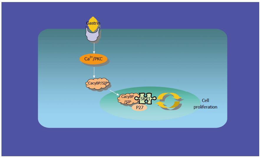
Figure 10 Interaction of gastrin, CacyBP/SIP, P27kip1 and Skp1 in gastric cancer.
- Citation: Niu YL, Li YJ, Wang JB, Lu YY, Liu ZX, Feng SS, Hu JG, Zhai HH. CacyBP/SIP nuclear translocation regulates p27Kip1 stability in gastric cancer cells. World J Gastroenterol 2016; 22(15): 3992-4001
- URL: https://www.wjgnet.com/1007-9327/full/v22/i15/3992.htm
- DOI: https://dx.doi.org/10.3748/wjg.v22.i15.3992


