Copyright
©The Author(s) 2016.
World J Gastroenterol. Apr 21, 2016; 22(15): 3885-3891
Published online Apr 21, 2016. doi: 10.3748/wjg.v22.i15.3885
Published online Apr 21, 2016. doi: 10.3748/wjg.v22.i15.3885
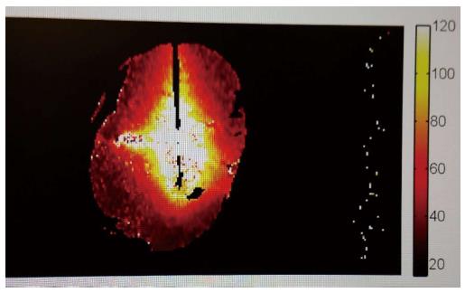
Figure 1 Thermal mapping using phase changes in magnetic resonance imaging, temperature code is depicted in the bar at the right margin of the image (values in degree Celsius).
Courtesy of MedWaves Inc., San Diego, CA, United States.
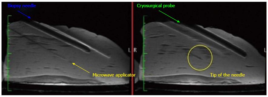
Figure 2 Magnetic resonance imaging of different devices for liver directed interventions.
Of note is the inaccuracy in displaying the position of the needle shaft and tip with older devices and the complete absence of artifacts with the use of a novel microwave applicator. Courtesy of MedWaves Inc., San Diego, CA, United States.
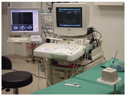
Figure 3 Clinical setup for ultrasound fusion imaging.
The ultrasound machine is visible with an additional monitor for displaying the previously digitally acquired cross-sectional examination images. Meanwhile, there are also systems with a split screen. The arrow points at the electromagnetic reference point.
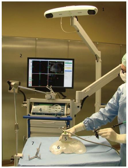
Figure 4 Example for a navigation device using optical tracking.
The crucial elements are shown under intraoperative conditions with a phantom liver model. 1: Stereooptic camera; 2: Monitor with a horizontally and vertically split screen; 3: Reference point; 4: Radiofrequency generator; 5: Ultrasound probe; 6: Liver phantom; 7: Pointer.
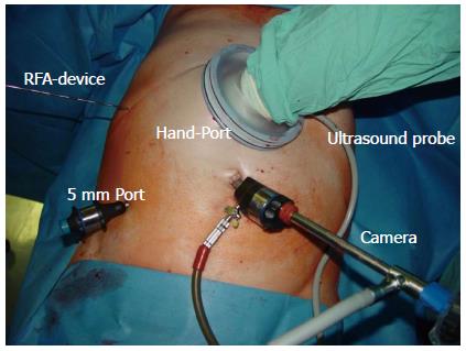
Figure 5 Clinical application of hand-assisted laparoscopic surgery.
Intraoperative radiofrequency ablation using hand-assistance. The advantages of a minimal-invasive approach are preserved.
- Citation: Eisele RM. Advances in local ablation of malignant liver lesions. World J Gastroenterol 2016; 22(15): 3885-3891
- URL: https://www.wjgnet.com/1007-9327/full/v22/i15/3885.htm
- DOI: https://dx.doi.org/10.3748/wjg.v22.i15.3885


