Copyright
©The Author(s) 2016.
World J Gastroenterol. Apr 14, 2016; 22(14): 3852-3859
Published online Apr 14, 2016. doi: 10.3748/wjg.v22.i14.3852
Published online Apr 14, 2016. doi: 10.3748/wjg.v22.i14.3852
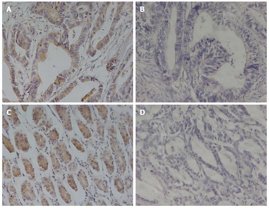
Figure 1 Comparison of immunohistochemical staining of αvβ6 and MMP-9 in the same gastric cancer specimen evaluated with formalin-fixed, paraffin-embedded tissue (streptavidin-peroxidase method; original magnification × 400).
Positive αvβ6 expression was detected predominantly in the cell membrane, and matrix metalloproteinase 9 (MMP-9) mainly exhibited a cytoplasmic immunostaining in tumor cells. A: Positive αvβ6 expression; B: Negative αvβ6 expression; C: Positive MMP-9 expression; D: Negative MMP-9 expression.
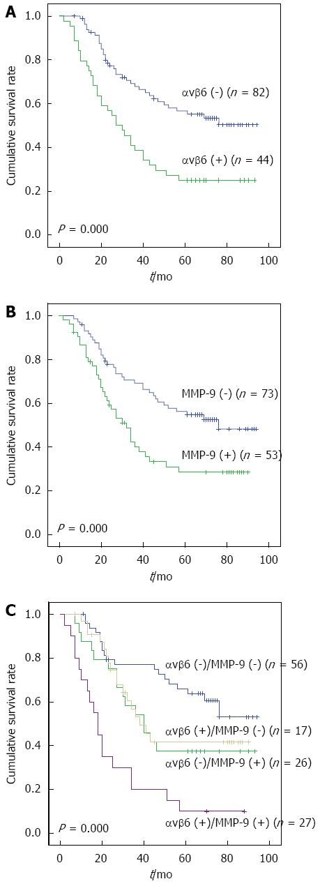
Figure 2 Kaplan-Meier plots show the association of survival and expression of integrin αvβ6 and MMP-9 in all gastric cancer patients tested.
A: Positive expression of αvβ6 was associated with decreased survival (P = 0.000); B: The same tendency was present in matrix metalloproteinase 9 (MMP-9) positive expression cases (P = 0.000); C: When αvβ6 and MMP-9 alterations were analyzed together, patients with both αvβ6 and MMP-9 positive expression experienced the highest mortality of the four groups (P = 0.000).
- Citation: Lian PL, Liu Z, Yang GY, Zhao R, Zhang ZY, Chen YG, Zhuang ZN, Xu KS. Integrin αvβ6 and matrix metalloproteinase 9 correlate with survival in gastric cancer. World J Gastroenterol 2016; 22(14): 3852-3859
- URL: https://www.wjgnet.com/1007-9327/full/v22/i14/3852.htm
- DOI: https://dx.doi.org/10.3748/wjg.v22.i14.3852


