Copyright
©The Author(s) 2016.
World J Gastroenterol. Mar 28, 2016; 22(12): 3432-3440
Published online Mar 28, 2016. doi: 10.3748/wjg.v22.i12.3432
Published online Mar 28, 2016. doi: 10.3748/wjg.v22.i12.3432
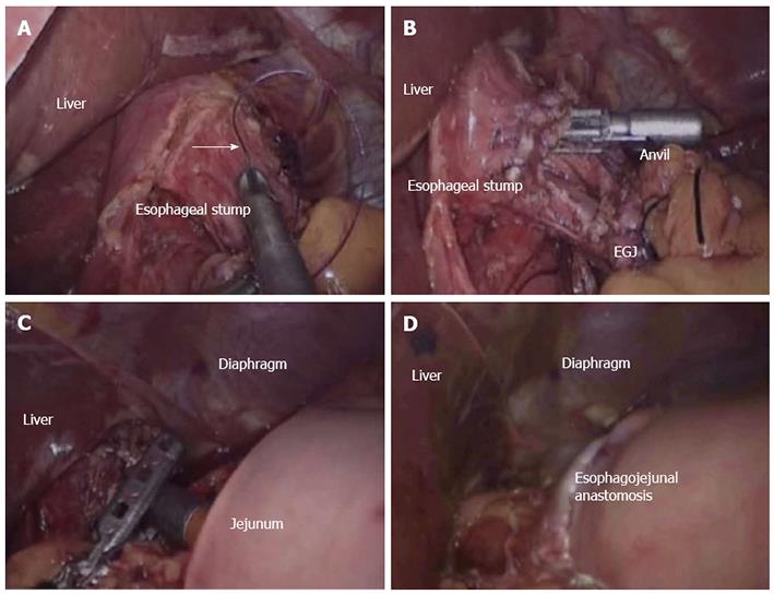
Figure 1 Conventional circular stapler-anvil method.
A: The purse-string suture (white arrow) was placed in the esophagus; B: The anvil was introduced into the esophageal stump through the hole; C: The circular stapler was introduced into the jejunum through the jejunal stump and attached with the anvil; D: The circular stapler was fired and the esophagojejunostomy was completed.
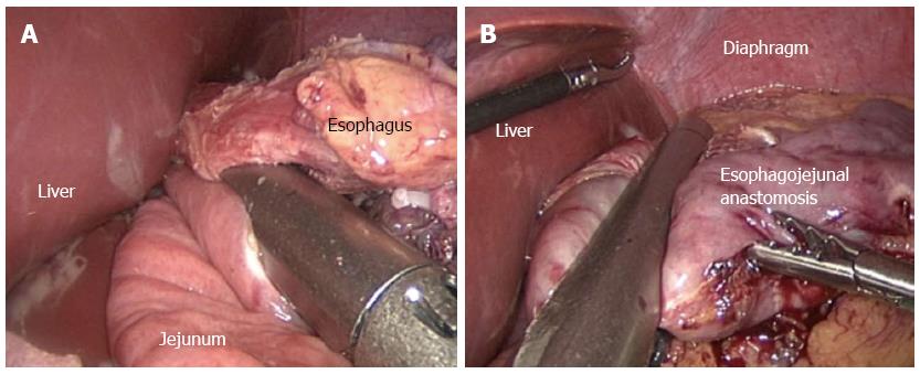
Figure 2 Linear stapler side-to-side method.
A: Each jaw of the linear stapler was inserted into the holes on the esophageal stump and the jejunum and then the linear stapler was fired; B: The entry hole and esophagus were closed using the stapler.
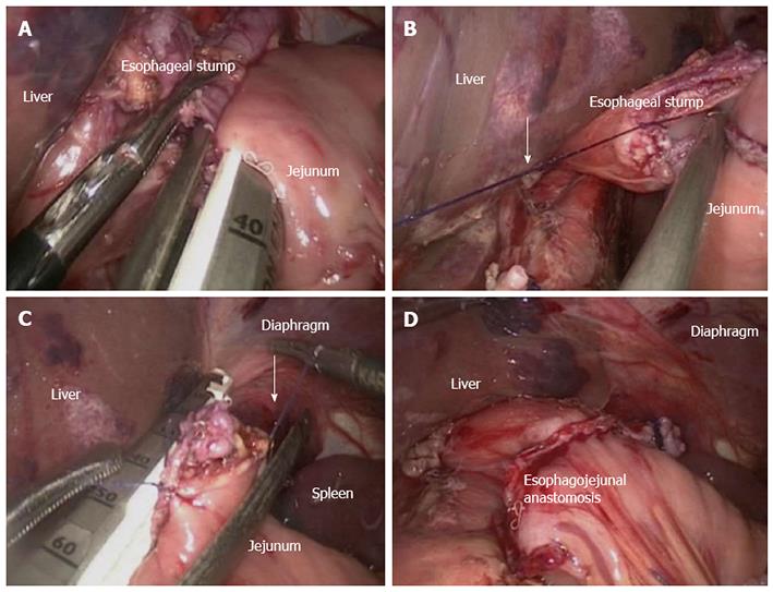
Figure 3 Linear stapler delta-shaped method.
A: Small holes were created along the edge of the esophageal stump and the jejunum that were approximated and joined with the endoscopic linear stapler; B: Stay sutures (white arrow) were placed to lift the common opening; C: The common opening was then closed with two applications of the linear stapler; D: Reconstruction of the intracorporeal alimentary tract was completed.
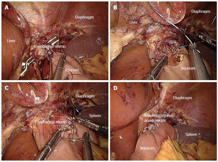
Figure 4 Hand-sewn end-to-side method.
A: The jejunum was anchored to the esophageal stump by several serosal muscularis interrupted sutures placed in the posterior layer of the esophageal stump; B: The posterior wall was closed by several full-thickness interrupted sutures; C: Closure of the anterior wall was carried out by a full-thickness continuous suture; D: The seromuscular layer was strengthened with interrupted sutures to reduce tension.
- Citation: Chen K, Pan Y, Cai JQ, Xu XW, Wu D, Yan JF, Chen RG, He Y, Mou YP. Intracorporeal esophagojejunostomy after totally laparoscopic total gastrectomy: A single-center 7-year experience. World J Gastroenterol 2016; 22(12): 3432-3440
- URL: https://www.wjgnet.com/1007-9327/full/v22/i12/3432.htm
- DOI: https://dx.doi.org/10.3748/wjg.v22.i12.3432


