Copyright
©The Author(s) 2016.
World J Gastroenterol. Mar 28, 2016; 22(12): 3335-3340
Published online Mar 28, 2016. doi: 10.3748/wjg.v22.i12.3335
Published online Mar 28, 2016. doi: 10.3748/wjg.v22.i12.3335
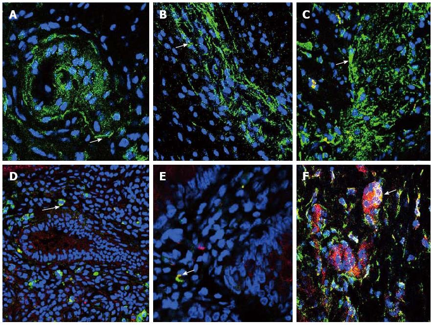
Figure 1 Immunofluorescent staining of platelet-derived growth factor receptor-α-positive-cells.
Immunofluorescent staining of platelet-derived growth factor receptor-α-positive (PDGFRα+)-cells (green) in the myenteric plexus of normal control (A), aganglionic (B) and ganglionic (C) colon, arrow shows cell body. Nuclei were stained with DAPI (blue). Mucosal PDGFRα+-cells (green) were seen to co-express TLR4 (red) (D) (arrow), TLR5 (red) (E) (arrow) and P2RY1 (red) (F) (arrow). A-F: Magnification × 40.
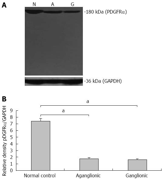
Figure 2 Western blot of platelet-derived growth factor receptor-α protein expression.
A: Platelet-derived growth factor receptor-α (PDGFRα) protein expression was high in normal controls and markedly decreased in both ganglionic and aganglionic specimens. The loading control GAPDH was similarly expressed in normal controls, ganglionic and aganglionic specimens; B: Densitometry analysis showed the range of PDGFRα protein expression among the samples studied. aP values are significant vs normal control.
- Citation: O’Donnell AM, Coyle D, Puri P. Deficiency of platelet-derived growth factor receptor-α-positive cells in Hirschsprung's disease colon. World J Gastroenterol 2016; 22(12): 3335-3340
- URL: https://www.wjgnet.com/1007-9327/full/v22/i12/3335.htm
- DOI: https://dx.doi.org/10.3748/wjg.v22.i12.3335


