Copyright
©The Author(s) 2016.
World J Gastroenterol. Mar 28, 2016; 22(12): 3305-3314
Published online Mar 28, 2016. doi: 10.3748/wjg.v22.i12.3305
Published online Mar 28, 2016. doi: 10.3748/wjg.v22.i12.3305
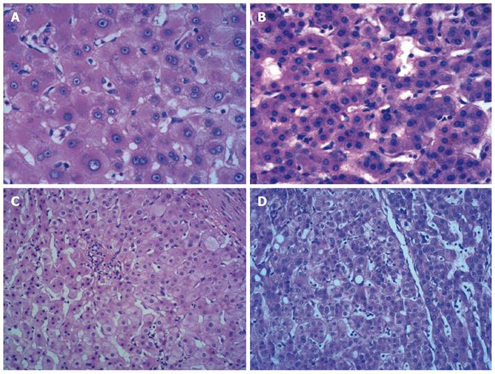
Figure 1 Histological characterization of large cell change (A), small cell change (B), low-grade dysplastic nodule (C) and high-grade dysplastic nodule (D).
Large cell change is characterized by cellular and nuclear enlargement with preserved nuclear and cytoplasmic ratio, nuclear pleomorphism and hyperchromasia, and prominent nucleoli (A), and small cell change characterized by decreased cell volume, mild nuclear pleomorphism, an increased nuclear-cytoplasmic ratio, and increased nuclear density (B); Low-grade dysplastic nodule is characterized by minimal cytological atypia, slightly increased nuclear-cytoplasmic ratio and cell density (C), and high-grade dysplastic nodule characterized by high cell density, an increased nuclear-cytoplasmic ratio, hepatocytes organized in trabeculae that are two to three cells thick, and mild nuclear atypia (D). A-D: Hematoxylin-eosin staining, magnification × 200.
- Citation: Niu ZS, Niu XJ, Wang WH, Zhao J. Latest developments in precancerous lesions of hepatocellular carcinoma. World J Gastroenterol 2016; 22(12): 3305-3314
- URL: https://www.wjgnet.com/1007-9327/full/v22/i12/3305.htm
- DOI: https://dx.doi.org/10.3748/wjg.v22.i12.3305


