Copyright
©The Author(s) 2016.
World J Gastroenterol. Mar 14, 2016; 22(10): 3062-3065
Published online Mar 14, 2016. doi: 10.3748/wjg.v22.i10.3062
Published online Mar 14, 2016. doi: 10.3748/wjg.v22.i10.3062
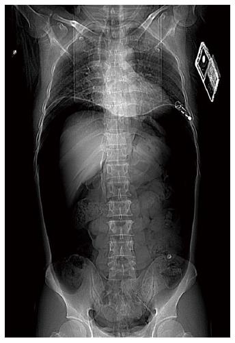
Figure 1 X-rays taken in the supine position, showing a large volume of air in the abdominal cavity, and elevated diaphragm.
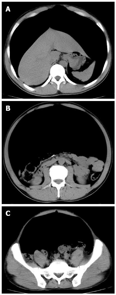
Figure 2 Computed tomography scan showing a large volume of air under the diaphragm (A), a large volume of air in the abdominal cavity (B) and in the pelvic cavity (C).
- Citation: Yin WB, Hu JL, Gao Y, Zhang XX, Zhang MS, Liu GW, Zheng XF, Lu Y. Rupture of sigmoid colon caused by compressed air. World J Gastroenterol 2016; 22(10): 3062-3065
- URL: https://www.wjgnet.com/1007-9327/full/v22/i10/3062.htm
- DOI: https://dx.doi.org/10.3748/wjg.v22.i10.3062


