Copyright
©The Author(s) 2015.
World J Gastroenterol. Mar 7, 2015; 21(9): 2848-2853
Published online Mar 7, 2015. doi: 10.3748/wjg.v21.i9.2848
Published online Mar 7, 2015. doi: 10.3748/wjg.v21.i9.2848
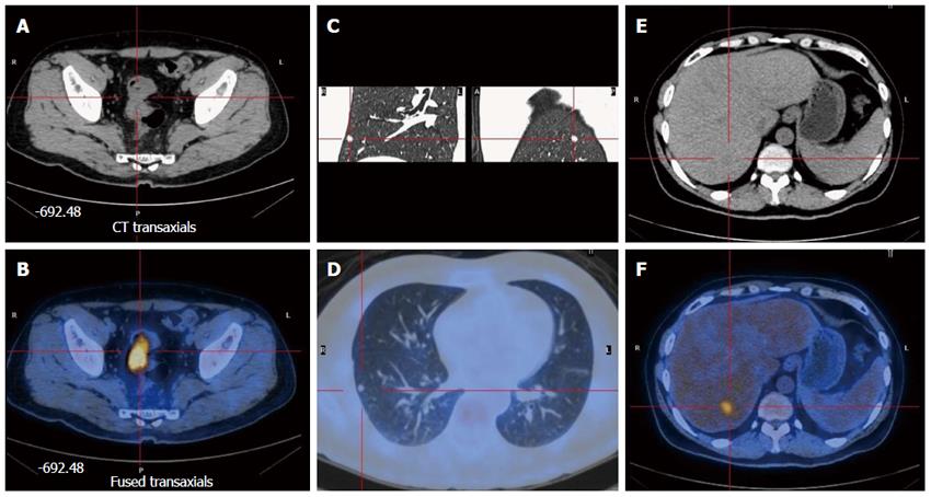
Figure 1 Preoperative positron emission tomography showed rectal cancer with liver and lung metastases.
A and B: Rectal cancer; C and D: Lung metastasis; E and F: Liver metastasis.
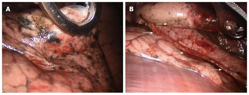
Figure 2 Robot-assisted wedge-shaped resection of the right lobe for lung metastasis.
A: Arrow indicates tumor location; B: Arrows indicate the cut line of the Endo GIA stapling device.
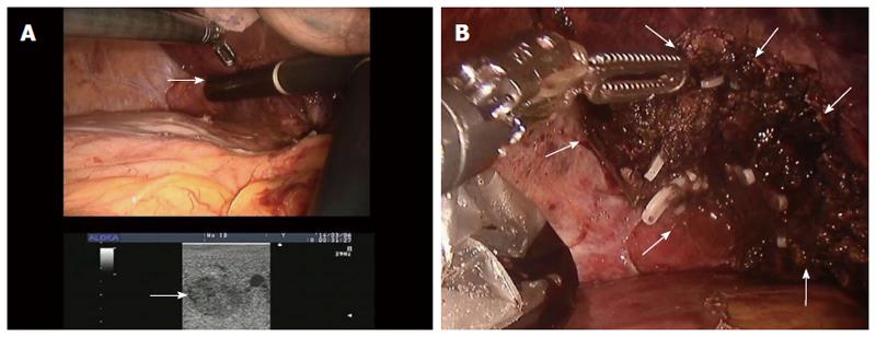
Figure 3 Robot-assisted segmental hepatectomy for liver metastasis.
A: Intraoperative ultrasound was used to mark tumor location; B: The tumor was resected.
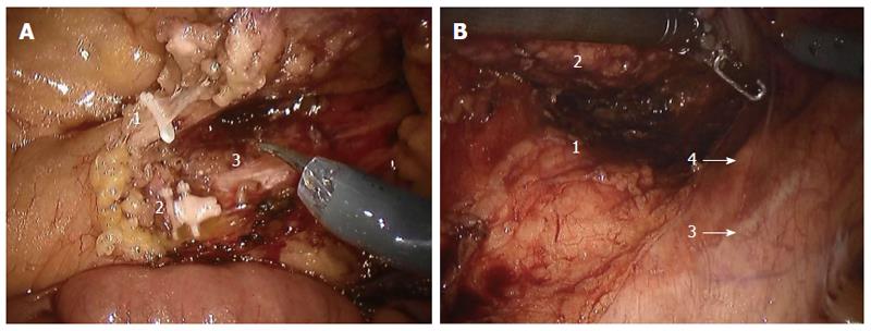
Figure 4 Operative images of robot-assisted anterior resection for rectal cancer.
A: 1, inferior mesenteric vein; 2, IMA; 3, inferior hypogastric nerve; B: 1, presacral space; 2, rectum; 3, iliac vessels; 4, ureter.
- Citation: Xu JM, Wei Y, Wang XY, Fan H, Chang WJ, Ren L, Jiang W, Fan J, Qin XY. Robot-assisted one-stage resection of rectal cancer with liver and lung metastases. World J Gastroenterol 2015; 21(9): 2848-2853
- URL: https://www.wjgnet.com/1007-9327/full/v21/i9/2848.htm
- DOI: https://dx.doi.org/10.3748/wjg.v21.i9.2848


