Copyright
©The Author(s) 2015.
World J Gastroenterol. Feb 28, 2015; 21(8): 2475-2482
Published online Feb 28, 2015. doi: 10.3748/wjg.v21.i8.2475
Published online Feb 28, 2015. doi: 10.3748/wjg.v21.i8.2475
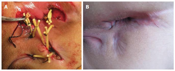
Figure 1 An 18-year-old male Crohn's disease patient.
A: A rubber band was placed during the surgery for drainage; B: Thirty weeks after the surgery.
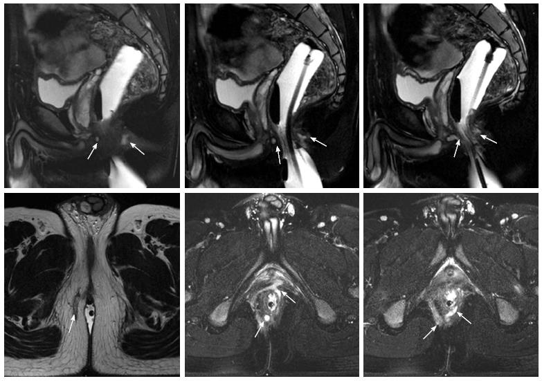
Figure 2 An 18-year-old male Crohn's disease patient, showing a full horseshoe transsphincteric anal fistula in magnetic resonance images (the rectum was extended by the intracavitary water cyst).
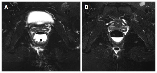
Figure 3 A 26-year-old female Crohn's disease patient.
A: Preoperative magnetic resonance imaging (MRI) suggested a transsphincteric anal fistula in the deep posterior space of the anal canal; B: Postoperative MRI showed that there was still a cavity within the posterior space of the anal canal (30 wk after surgery). No obvious clinical symptoms were found when the patient was followed for 34 mo (the rectum was extended by the intracavitary water cyst).
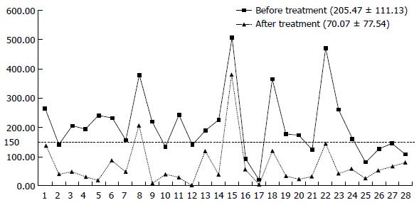
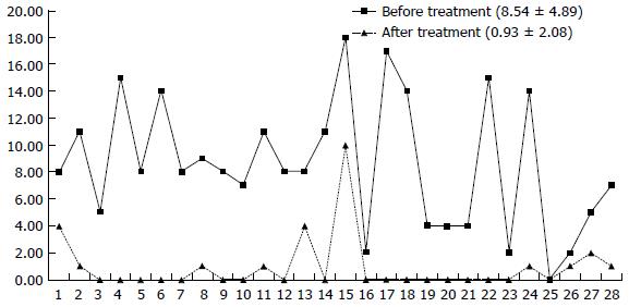
Figure 4 Crohn's disease activity index results before and after treatment.
Figure 5 Perianal Crohn's disease activity index results before and after treatment.
- Citation: Yang BL, Chen YG, Gu YF, Chen HJ, Sun GD, Zhu P, Shao WJ. Long-term outcome of infliximab combined with surgery for perianal fistulizing Crohn's disease. World J Gastroenterol 2015; 21(8): 2475-2482
- URL: https://www.wjgnet.com/1007-9327/full/v21/i8/2475.htm
- DOI: https://dx.doi.org/10.3748/wjg.v21.i8.2475


