Copyright
©The Author(s) 2015.
World J Gastroenterol. Feb 28, 2015; 21(8): 2467-2474
Published online Feb 28, 2015. doi: 10.3748/wjg.v21.i8.2467
Published online Feb 28, 2015. doi: 10.3748/wjg.v21.i8.2467
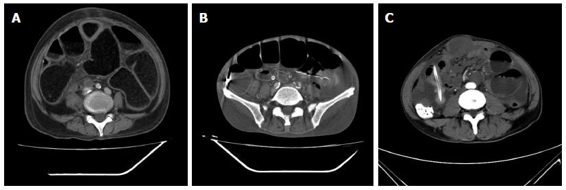
Figure 1 Computed tomography findings of small bowel obstruction before the procedure.
A: Significantly dilated intestines with local wall thickening; B: Accumulated gas in the dilated intestinal cavity, with visible fluid level; C: Local intestinal dilatation, partial bowel and abdominal wall adhesions, and a substantial mass is visible.
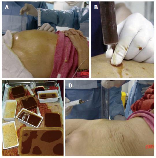
Figure 2 Procedural images involving a 75-year-old man with recurrent gastric cancer.
A: Preoperative abdominal distension and skin tension; B and C: A large amount of liquid was extracted intraoperatively (700 mL gas and liquid 2800 mL liquid); D: Abdomen returned to normal and the skin was relaxed.
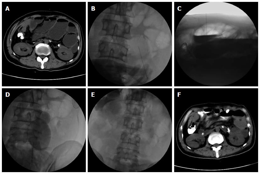
Figure 3 Computed tomography findings in a 75-year-old man with recurrent gastric cancer.
A: Preoperative computed tomography (CT) showed significant dilation with visible gas and effusions; B-D: Anteroposterior radiographs during the percutaneous procedure and suction under imaging guidance; E: At the end of aspiration, gas and effusions were significantly reduced; F: Postoperative CT showed that the intestine had returned to normal.
- Citation: Jiang TH, Sun XJ, Chen Y, Cheng HQ, Fang SM, Jiang HS, Cao Y, Liu BY, Wu SQ, Mao AW. Percutaneous needle decompression in treatment of malignant small bowel obstruction. World J Gastroenterol 2015; 21(8): 2467-2474
- URL: https://www.wjgnet.com/1007-9327/full/v21/i8/2467.htm
- DOI: https://dx.doi.org/10.3748/wjg.v21.i8.2467


