Copyright
©The Author(s) 2015.
World J Gastroenterol. Feb 14, 2015; 21(6): 2000-2004
Published online Feb 14, 2015. doi: 10.3748/wjg.v21.i6.2000
Published online Feb 14, 2015. doi: 10.3748/wjg.v21.i6.2000
Figure 1 Imaging of the intra- and extrahepatic bile ducts.
A: Magnetic resonance cholangiopancreatography showing a large number of bile duct stones within the intra- and extrahepatic bile ducts (arrows) with a mild dilation of the intrahepatic bile duct; B: Percutaneous transhepatic cholangiography showing the bile duct cast, which is a result of molding of the bile ducts by the stones (arrows).
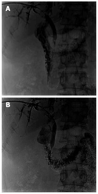
Figure 2 Crushing of the extrahepatic bile duct stones.
Percutaneous transhepatic cholangiography showing that bile duct stones were crushed from the A: Proximal; B: Distal duct with the inflated balloon catheter.
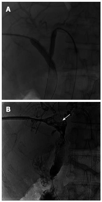
Figure 3 Percutaneous interventional techniques used on the intrahepatic bile stones.
A: The intrahepatic bile stones were crushed with the double balloon catheter technique; B: The large residual stones were cut with the basket extractor (arrow).
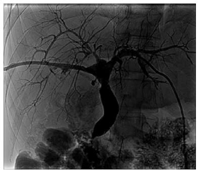
Figure 4 Elimination of bile duct stones.
Percutaneous transhepatic cholangiography showed no presence of the bile stones and a normal biliary tree four months after the percutaneous interventional procedures.
- Citation: Zhou CG, Wei BJ, Gao K, Dai DK, Zhai RY. Successful treatment of complex cholangiolithiasis following orthotopic liver transplantation with interventional radiology. World J Gastroenterol 2015; 21(6): 2000-2004
- URL: https://www.wjgnet.com/1007-9327/full/v21/i6/2000.htm
- DOI: https://dx.doi.org/10.3748/wjg.v21.i6.2000


