Copyright
©The Author(s) 2015.
World J Gastroenterol. Dec 28, 2015; 21(48): 13524-13531
Published online Dec 28, 2015. doi: 10.3748/wjg.v21.i48.13524
Published online Dec 28, 2015. doi: 10.3748/wjg.v21.i48.13524
Figure 1 Dynamic enhancing pattern of the liver metastasis from hepatoid adenocarcinoma of the stomach in a 63-yr-old male.
A: Liver metastases from hepatoid adenocarcinoma of the stomach presented with hypoattenuation on precontrast computed tomography (CT); B and C: The nodules revealed arterial hyperattenuation (B) and late phase washout (C) on dynamic CT study. Central necrosis (arrows) was found in the small liver nodules; D: The patient received transarterial chemoembolization for liver metastases. The treated liver nodules were well embolized by densely packed Lipiodol (arrows). However, new liver metastases were observed on follow-up CT. Pre: Precontrast; A-phase: Arterial phase; PV-phase: Portal venous phase; F/U: Follow-up image.
Figure 2 Dynamic enhancing pattern of the liver metastasis from hepatoid adenocarcinoma of the stomach in a 69-yr-old male.
A: Liver metastasis from hepatoid adenocarcinoma of the stomach presented with hypoattenuation on precontrast computed tomography (CT); B and C: The liver mass revealed arterial hyperattenuation (B) and late phase washout (C) on dynamic CT study. Central necrosis and tumor hemorrhage (arrow) were noted; D: The patient received total gastrectomy combined with systemic chemotherapy. Progression of the liver metastasis with direct IVC invasion (arrow) was observed on follow-up CT. Pre: Precontrast; A-phase: Arterial phase; PV-phase: Portal venous phase; F/U: Follow-up image.
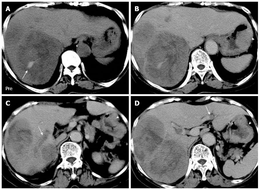
Figure 3 Venous tumor thrombosis in a 69-yr-old female with hepatoid adenocarcinoma of the stomach and liver metastases.
A: Bulky liver metastases presented with tumor hemorrhage (arrow) on precontrast computed tomography; B and C: Eccentric wall thickening and heterogenous enhancement of the gastric body (arrowheads) implied gastric malignancy. On portal venous phase, the liver mass presented irregular central necrosis (B) and right portal vein tumor thrombosis (arrow; C); D: Isolated left portal vein tumor thrombosis (arrow) was observed. No visible liver nodule was found at the left liver lobe. Pre: Precontrast.
- Citation: Lin YY, Chen CM, Huang YH, Lin CY, Chu SY, Hsu MY, Pan KT, Tseng JH. Liver metastasis from hepatoid adenocarcinoma of the stomach mimicking hepatocellular carcinoma: Dynamic computed tomography findings. World J Gastroenterol 2015; 21(48): 13524-13531
- URL: https://www.wjgnet.com/1007-9327/full/v21/i48/13524.htm
- DOI: https://dx.doi.org/10.3748/wjg.v21.i48.13524


