Copyright
©The Author(s) 2015.
World J Gastroenterol. Dec 21, 2015; 21(47): 13240-13249
Published online Dec 21, 2015. doi: 10.3748/wjg.v21.i47.13240
Published online Dec 21, 2015. doi: 10.3748/wjg.v21.i47.13240
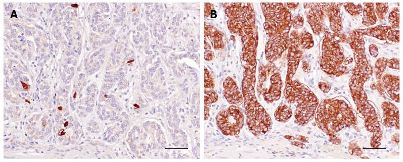
Figure 1 Histidine decarboxylase (A) and vesicular monoamine transporter 2 (B) immunostained type I enterochromaffin-like cell neuroendocrine tumour (consecutive sections).
Only a few scattered histidine decarboxylase (HDC)-immunoreactive cells are present in the tumour, whereas virtually all neoplastic cells display VMAT-2 immunoreactivity. Bars = 50 μm (A, B).
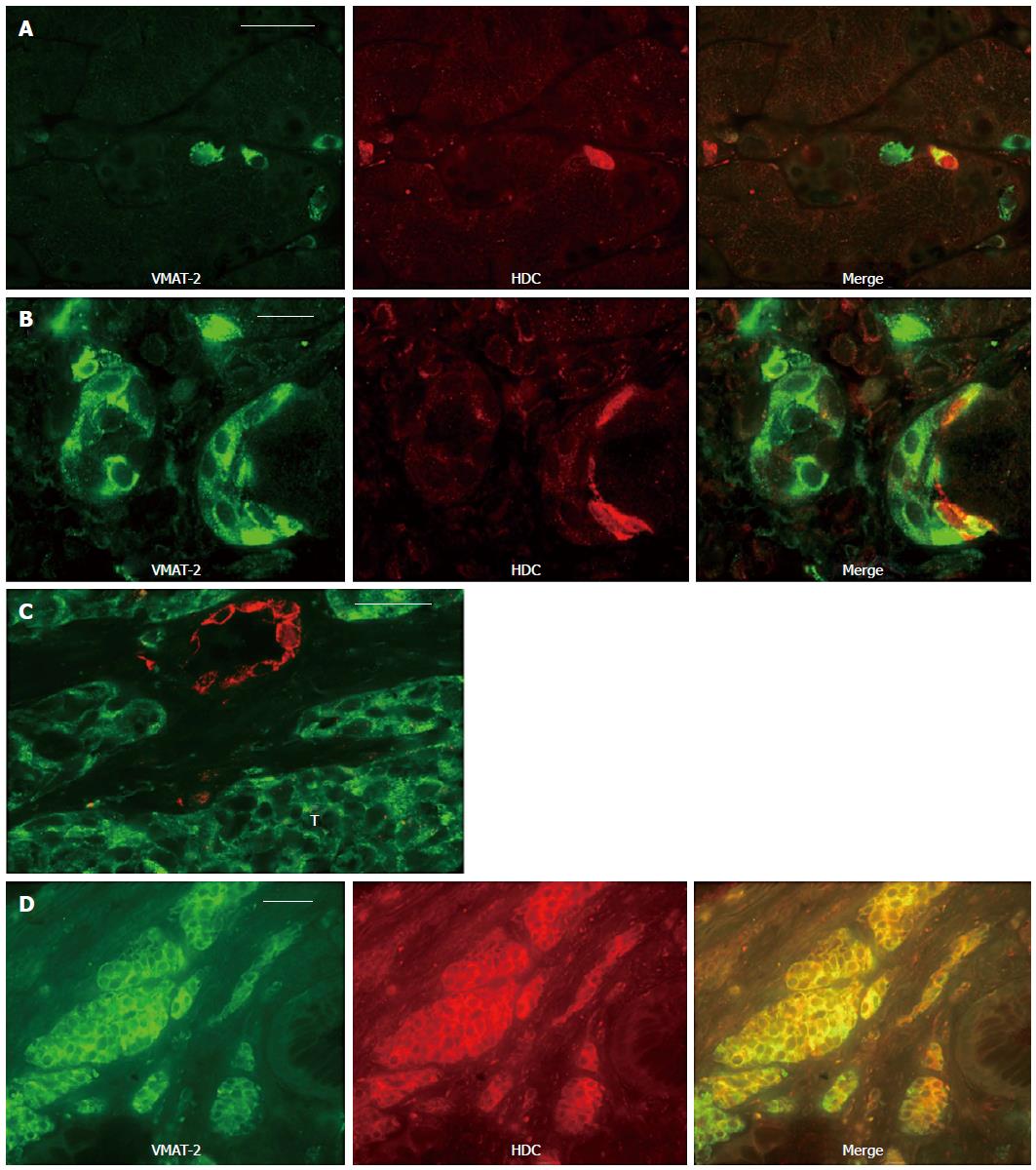
Figure 2 Co-localization studies.
A: Normal human oxyntic mucosa double-immunostained for vesicular monoamine transporter 2 (VMAT-2) (green) and histidine decarboxylase (HDC) (red). Co-localization (yellow) depicts that VMAT-2 and HDC are co-expressed in the same neuroendocrine cell, whereas the remaining immunoreactive (IR) cells in the gastric glands do not display co-expression; B: Oxyntic mucosa adjacent to a type I enterochromaffin-like (ECL) cell neuroendocrine tumour (NET) double immunostained for VMAT-2 (green) and HDC (red). In one focus of neuroendocrine cell hyperplasia identified by VMAT-2 (linear pattern), only a fraction of cells co-express HDC (yellow), whereas another is non-IR for the enzyme; C: Oxyntic mucosa adjacent to a type I ECL cell NET double immunostained for VMAT-2 (green) and HDC (red). The HDC-IR cells display a linear pattern of neuroendocrine cell hyperplasia and do not co-express the transporter protein. In the foci of nodular cell hyperplasia identified by VMAT-2, no cell is HDC-IR. Adjacent tumour cells (T) reveal only VMAT-2 immunoreactivity; D: A malignant type III ECL cell NET accompanied by increased U-MeImAA excretion was double-immunostained for VMAT-2 (green) and HDC (red). Co-localization (yellow) shows that the tumour cells that are HDC-IR co-express the transporter protein. VMAT-2-IR cells slightly outnumber the HDC-IR cells. Bars = 30 μm (A, C, D); 20 μm (B).
- Citation: Tsolakis AV, Grimelius L, Granerus G, Stridsberg M, Falkmer SE, Janson ET. Histidine decarboxylase and urinary methylimidazoleacetic acid in gastric neuroendocrine cells and tumours. World J Gastroenterol 2015; 21(47): 13240-13249
- URL: https://www.wjgnet.com/1007-9327/full/v21/i47/13240.htm
- DOI: https://dx.doi.org/10.3748/wjg.v21.i47.13240


