Copyright
©The Author(s) 2015.
World J Gastroenterol. Sep 21, 2015; 21(35): 10246-10250
Published online Sep 21, 2015. doi: 10.3748/wjg.v21.i35.10246
Published online Sep 21, 2015. doi: 10.3748/wjg.v21.i35.10246
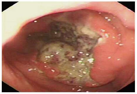
Figure 1 Endoscopic imaging of the gastric tumor.
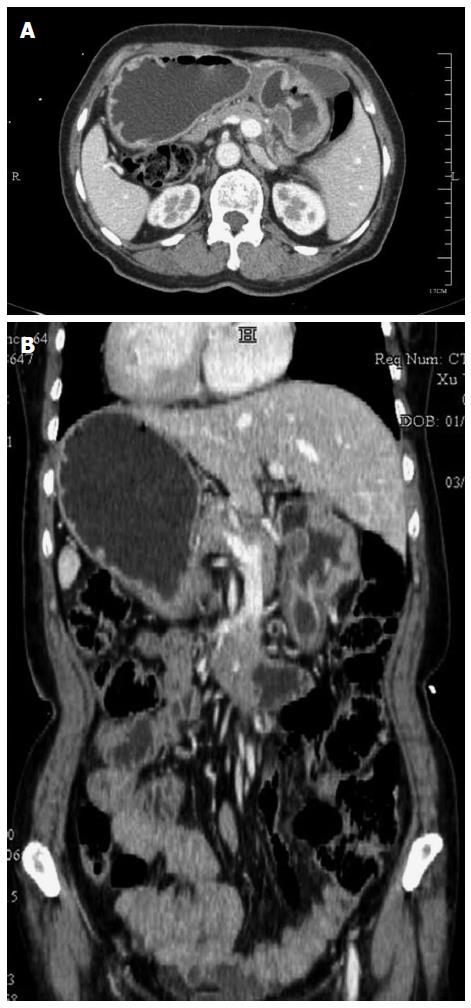
Figure 2 Enhanced abdominal computed tomography imaging.
A: Computed tomography (CT) scan revealing thickening of the wall of the antrum, without enlarged lymph nodes. B: CT scan showing that all of the organs inside of the abdomen are inversely positioned.
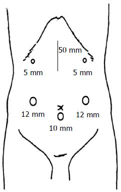
Figure 3 Sites of trocar placement.
A camera was inserted into the subumbilical area through a 10-mm trocar.
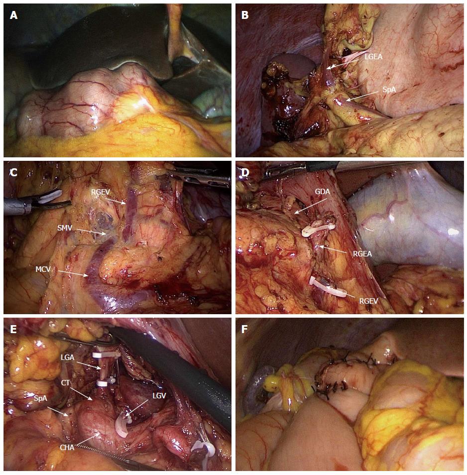
Figure 4 Intraoperative findings.
A: Laparoscopic view showing inversion of the intraabdominal organs. B: The left gastroepiploic artery located on the right side was exposed. C: After exposure of the root of the middle colic vein and superior mesenteric vein, station no. 14v lymph node dissection was completed. D: The right gastroepiploic artery located on the left side, bifurcating from the gastroduodenal artery, was ligated. E: The left gastric artery located on the right side was ligated, allowing for dissection of station nos. 7 and 9 lymph nodes. F: Billroth II reconstruction was performed. SpA: Splenic artery; LGEA: Left gastroepiploic artery; SMV: Superior mesenteric vein; MCV: Middle colic vein; RGEV: Right gastroepiploic vein; RGEA: Right gastroepiploic artery; GDA: Gastroduodenal artery; CHA: Common hepatic artery; LGA: Left gastric artery; LGV: Left gastric vein; CT: Celiac trunk.
- Citation: Ye MF, Tao F, Xu GG, Sun AJ. Laparoscopy-assisted distal gastrectomy for advanced gastric cancer with situs inversus totalis: A case report. World J Gastroenterol 2015; 21(35): 10246-10250
- URL: https://www.wjgnet.com/1007-9327/full/v21/i35/10246.htm
- DOI: https://dx.doi.org/10.3748/wjg.v21.i35.10246


