Copyright
©The Author(s) 2015.
World J Gastroenterol. Sep 21, 2015; 21(35): 10184-10191
Published online Sep 21, 2015. doi: 10.3748/wjg.v21.i35.10184
Published online Sep 21, 2015. doi: 10.3748/wjg.v21.i35.10184
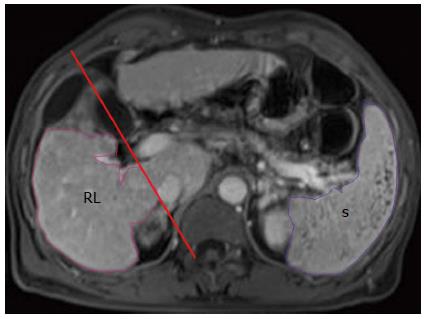
Figure 1 Outlines of right liver lobe (RL, in pink) and the spleen (S, in purple) are delineated on the axial enhanced magnetic resonance image.
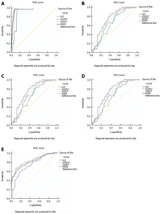
Figure 2 Right liver lobe volume × platelet count/spleen volume is better than platelet count, spleen volume/platelet count or spleen volume index/platelet count for distinguishing cirrhotic patients from healthy participants (A), Child-Pugh class A of cirrhosis from B, A from C, and B from C; and SV/PLT is better than PLT, RVPS or SI/PLT for identifying esophageal varices (E).
RVPS: Right liver lobe volume × platelet count/spleen volume; PLT: Platelet count; SV/PLT: Spleen volume/platelet count; SI/PLT: Spleen volume index/platelet count.
- Citation: Chen XL, Chen TW, Zhang XM, Li ZL, Zeng NL, Zhou P, Li H, Ren J, Xu GH, Hu JN. Platelet count combined with right liver volume and spleen volume measured by magnetic resonance imaging for identifying cirrhosis and esophageal varices. World J Gastroenterol 2015; 21(35): 10184-10191
- URL: https://www.wjgnet.com/1007-9327/full/v21/i35/10184.htm
- DOI: https://dx.doi.org/10.3748/wjg.v21.i35.10184


