Copyright
©The Author(s) 2015.
World J Gastroenterol. Aug 21, 2015; 21(31): 9387-9393
Published online Aug 21, 2015. doi: 10.3748/wjg.v21.i31.9387
Published online Aug 21, 2015. doi: 10.3748/wjg.v21.i31.9387
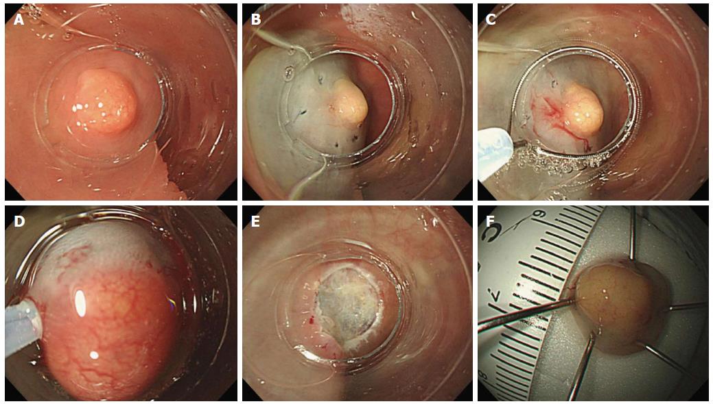
Figure 1 Endoscopic mucosal resection with a cap.
A: Transparent cap is attached to the distal end of the scope; B: Saline solution with indigo-carmine solution is injected submucosally beneath the tumor; C: A crescent-shaped snare is positioned on the internal circumferential ridge; D: The submucosal layer is suctioned and dissected with the snare; E: A clear resection surface is observed; F: The resection specimen is retrieved and measured. EMR-C: Endoscopic mucosal resection with a cap.
- Citation: Park SB, Kim HW, Kang DH, Choi CW, Kim SJ, Nam HS. Advantage of endoscopic mucosal resection with a cap for rectal neuroendocrine tumors. World J Gastroenterol 2015; 21(31): 9387-9393
- URL: https://www.wjgnet.com/1007-9327/full/v21/i31/9387.htm
- DOI: https://dx.doi.org/10.3748/wjg.v21.i31.9387


