Copyright
©The Author(s) 2015.
World J Gastroenterol. Aug 14, 2015; 21(30): 9228-9232
Published online Aug 14, 2015. doi: 10.3748/wjg.v21.i30.9228
Published online Aug 14, 2015. doi: 10.3748/wjg.v21.i30.9228
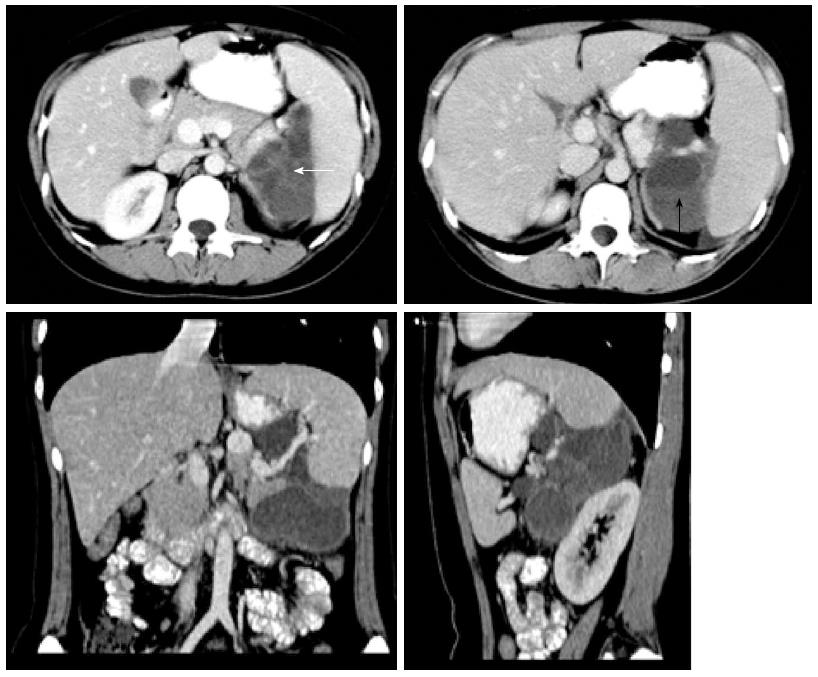
Figure 1 Enhanced computed tomography images of a 28-year-old female with pancreatic hemangioma.
The tumor was a multilocular cyst with septa (left; white arrow) and fluid-fluid levels (right; black arrow).
- Citation: Lu T, Yang C. Rare case of adult pancreatic hemangioma and review of the literature. World J Gastroenterol 2015; 21(30): 9228-9232
- URL: https://www.wjgnet.com/1007-9327/full/v21/i30/9228.htm
- DOI: https://dx.doi.org/10.3748/wjg.v21.i30.9228


