Copyright
©The Author(s) 2015.
World J Gastroenterol. Aug 14, 2015; 21(30): 9126-9133
Published online Aug 14, 2015. doi: 10.3748/wjg.v21.i30.9126
Published online Aug 14, 2015. doi: 10.3748/wjg.v21.i30.9126
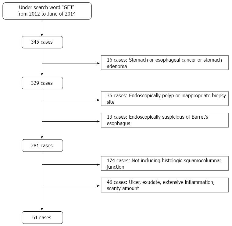
Figure 1 Patient and tissue selection.
All included samples were obtained from the gastroesophageal junction (GEJ) with histologic squamocolumnar junctions and proper amounts of glandular components.
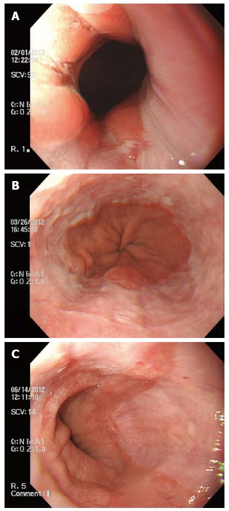
Figure 2 Endoscopic findings of gastroesophageal junction.
A: Los Angeles (LA) classification 0 indicates normal gastroesophageal junction (GEJ) with no mucosal breaks; this case showed oxyntic mucosa at the GEJ histologically; B: LA classification A indicating one or more mucosal breaks ≤ 5 mm in length. Microscopically, it showed cardiac mucosa at the GEJ; C: LA classification B with one or more mucosal breaks > 5 mm in length; this case had cardiac mucosa at the GEJ.
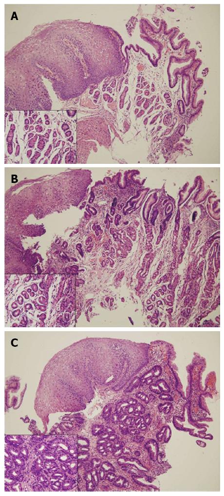
Figure 3 Columnar epithelium of squamocolumnar junction.
A: Oxyntic mucosa composed entirely of parietal and chief cells without mucous cells below the foveolar region; B: Oxyntocardiac mucosa containing a mixture of mucous cells and parietal cells; C: Cardiac mucosa composed entirely of mucous cells without any parietal cells (hematoxylin-eosin staining, × 100 magnification). Insets show high magnification finding (× 400 magnification) of glandular component of the gastroesophageal junction.
- Citation: Kim A, Park WY, Shin N, Lee HJ, Kim YK, Lee SJ, Hwang CS, Park DY, Kim GH, Lee BE, Jo HJ. Cardiac mucosa at the gastroesophageal junction: An Eastern perspective. World J Gastroenterol 2015; 21(30): 9126-9133
- URL: https://www.wjgnet.com/1007-9327/full/v21/i30/9126.htm
- DOI: https://dx.doi.org/10.3748/wjg.v21.i30.9126


