Copyright
©The Author(s) 2015.
World J Gastroenterol. Aug 7, 2015; 21(29): 8943-8951
Published online Aug 7, 2015. doi: 10.3748/wjg.v21.i29.8943
Published online Aug 7, 2015. doi: 10.3748/wjg.v21.i29.8943
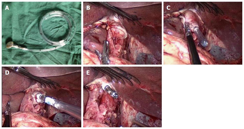
Figure 1 OrVilTM procedure.
A: The central rod of the anvil connected with a tube; B: The lower esophagus was dissociated, and the esophagus was closed and cut; C: The tube of the OrVilTM system was transorally inserted into the esophagus, and the head of the tube was pulled out from the small hole at the end of the esophagus; D: The tube was pulled out until the anvil that connected with the end of the tube reached the esophageal stump; E: The connection line between the tube and anvil was cut down, and the tube was pulled out.
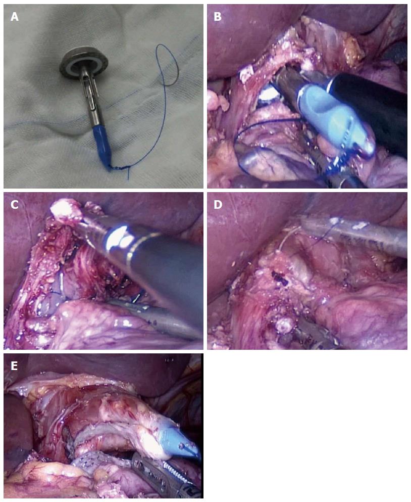
Figure 2 Hemi-double stapling technique procedure.
A: The tail of the central rod of the anvil was sutured with sutures that contained a needle; B: The anvil with needle-containing sutures was inserted into the esophagus via the incision on the lower edge of the esophagus; C: The needle was inserted through the anterior wall of the esophagus, and the needle and sutures were pulled out from the anterior wall of the esophagus; D: The lower edge of the esophagus was closed off and cut down using a linear cutter; E: The sutures were further retracted until the central rod of the anvil was completely pulled out.
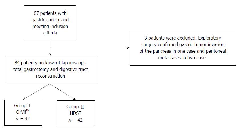
Figure 3 Patients’ flowchart.
Flowchart of the subject inclusion and allocation into group I and group II for the different insertion techniques for esophagojejunostomy. HDST: Hemi-double stapling technique.
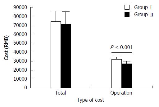
Figure 4 Comparison of the total cost of hospitalization and operation between the two groups.
Dark grey represents the cost using the OrVilTM system (Group I) and light grey represents the cost using the hemi-double stapling technique (HDST) method (Group II). The cost of operation using the OrVilTM system was significantly greater than that of using the HDST method (P < 0.001, Group I vs Group II).
- Citation: Wang H, Hao Q, Wang M, Feng M, Wang F, Kang X, Guan WX. Esophagojejunostomy after laparoscopic total gastrectomy by OrVilTM or hemi-double stapling technique. World J Gastroenterol 2015; 21(29): 8943-8951
- URL: https://www.wjgnet.com/1007-9327/full/v21/i29/8943.htm
- DOI: https://dx.doi.org/10.3748/wjg.v21.i29.8943


