Copyright
©The Author(s) 2015.
World J Gastroenterol. Jun 28, 2015; 21(24): 7553-7557
Published online Jun 28, 2015. doi: 10.3748/wjg.v21.i24.7553
Published online Jun 28, 2015. doi: 10.3748/wjg.v21.i24.7553
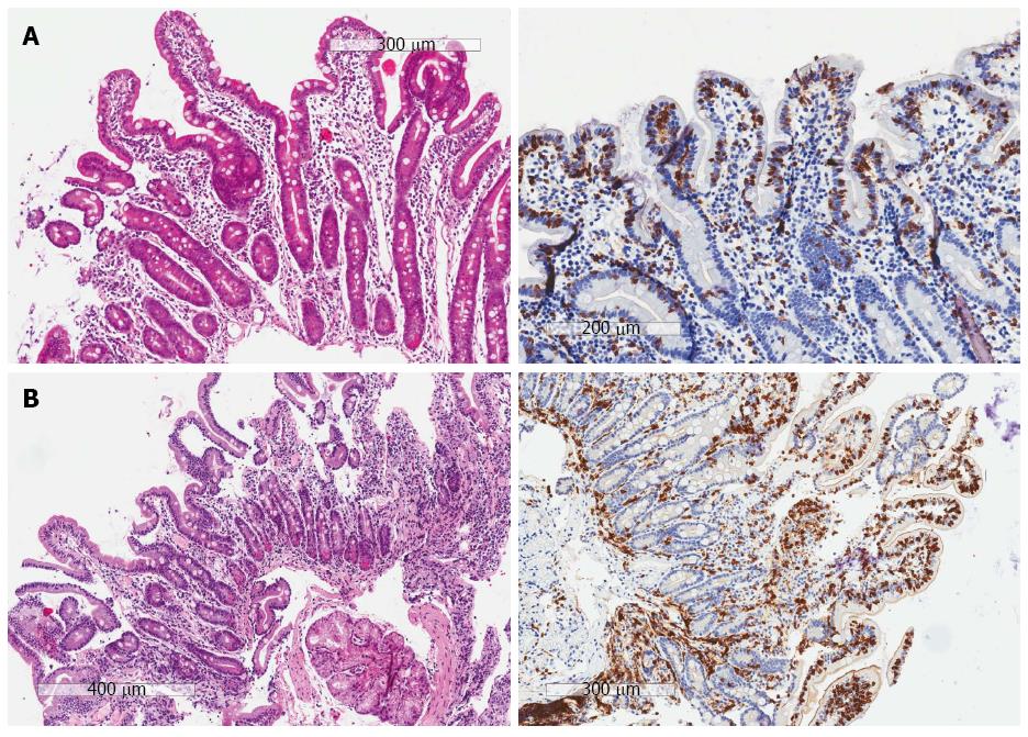
Figure 1 Examples of histological hematoxylin and eosin and CD3 sections.
A: The HE sections (left) show partial villous atrophy and crypt hyperplasia, but no intra-epithelial lymphocytosis, thus the HE sections do not reveal celiac disease. However, on the CD3 sections (right) the intra-epithelial lymphocytosis becomes clear and the diagnosis of celiac disease can be made (Marsh IIIa). The patient had clinical symptoms associated with celiac disease, positive serology and also responded well to the diet. The final diagnosis is celiac disease; B: The HE sections (left) show a Marsh 0, while the CD3 sections (right) show a Marsh I lesion. The patient had clinical symptoms associated with celiac disease, positive serology and also responded well to the diet. The final diagnosis is Marsh I.
- Citation: Mubarak A, Wolters VM, Houwen RH, ten Kate FJ. Immunohistochemical CD3 staining detects additional patients with celiac disease. World J Gastroenterol 2015; 21(24): 7553-7557
- URL: https://www.wjgnet.com/1007-9327/full/v21/i24/7553.htm
- DOI: https://dx.doi.org/10.3748/wjg.v21.i24.7553


