Copyright
©The Author(s) 2015.
World J Gastroenterol. May 28, 2015; 21(20): 6271-6279
Published online May 28, 2015. doi: 10.3748/wjg.v21.i20.6271
Published online May 28, 2015. doi: 10.3748/wjg.v21.i20.6271
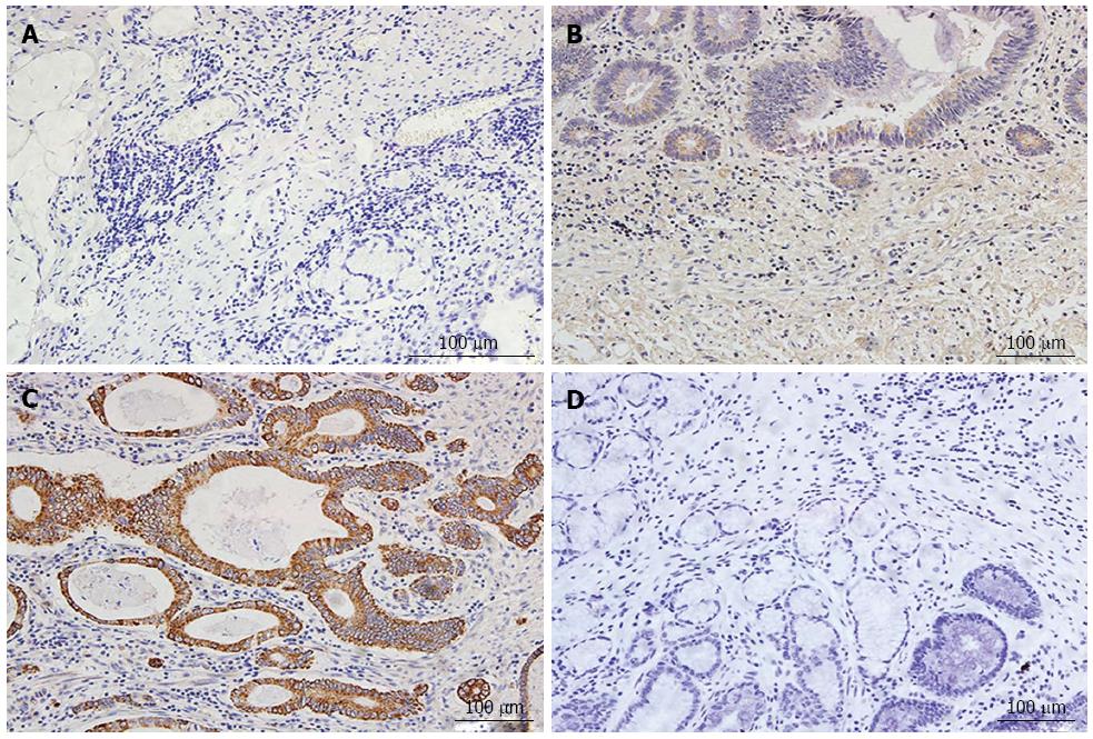
Figure 1 Immunohistochemical staining results for NADPH oxidase 2.
A: Negative PBS control in gastric cancer; B: Weak expression of NADPH oxidase 2 (NOX2) in gastric cancer, C: Strongly positive expression of NOX2 in gastric cancer, D: Negative expression of NOX2 in adjacent noncancerous tissue (× 200).
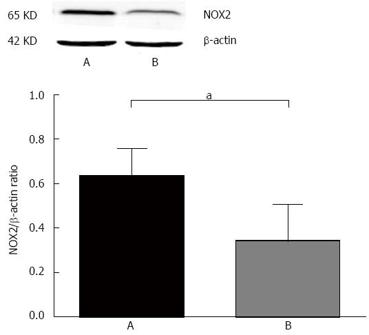
Figure 2 Western blot results for NADPH oxidase 2.
A: Gastric cancer tissue; B: Adjacent noncancerous tissue. aP < 0.05 vs group A.
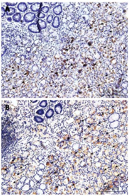
Figure 3 Localization of NOX2 and CD68 staining.
A: NOX2 expression in gastric cancer; B: CD68 expression in gastric cancer (× 200).
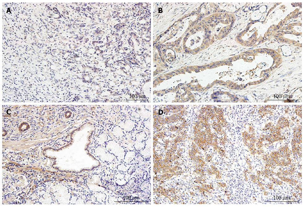
Figure 4 Immuohistochemistry staining results for vascular endothelial growth factor and epidermal growth factor receptor in gastric cancer.
A: Weakly positive expression of vascular endothelial growth factor (VEGF) in gastric cancer; B: Strongly positive expression of VEGF in gastric cancer; C: Weakly positive expression of epidermal growth factor receptor (EGFR) in gastric cancer; D: Strongly positive expression of EGFR in gastric cancer (× 200).
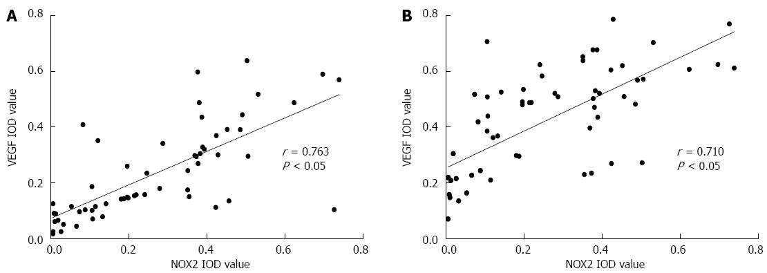
Figure 5 Expression of NADPH oxidase 2 was positively correlated with vascular endothelial growth factor and epidermal growth factor receptor in gastric cancer.
A: Spearman correlation between NOX2 and vascular endothelial growth factor; B: Spearman correlation between NOX2 and epidermal growth factor receptor. VEGF: Vascular endothelial growth factor; EGFR: Epidermal growth factor receptor.
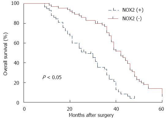
Figure 6 Follow-up investigation of the 5-year survival study showed that the patients who were NADPH oxidase 2 positive clearly presented a worse outcome than the NADPH oxidase 2 negative patients.
- Citation: Wang P, Shi Q, Deng WH, Yu J, Zuo T, Mei FC, Wang WX. Relationship between expression of NADPH oxidase 2 and invasion and prognosis of human gastric cancer. World J Gastroenterol 2015; 21(20): 6271-6279
- URL: https://www.wjgnet.com/1007-9327/full/v21/i20/6271.htm
- DOI: https://dx.doi.org/10.3748/wjg.v21.i20.6271


