Copyright
©2014 Baishideng Publishing Group Co.
World J Gastroenterol. Feb 28, 2014; 20(8): 2113-2116
Published online Feb 28, 2014. doi: 10.3748/wjg.v20.i8.2113
Published online Feb 28, 2014. doi: 10.3748/wjg.v20.i8.2113
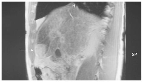
Figure 1 Magnetic resonance imaging showed a tumor-like lesion (arrow) and ischemia in segment 4 of the liver.
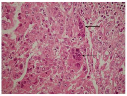
Figure 2 Hematoxylin and eosin-staining of a representative hepatocellular carcinoma section showing the multinuclear giant syncytial cells (arrows).
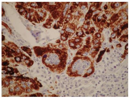
Figure 3 Mononuclear and multinuclear carcinoma cells showed reactivity for the hepatocyte antigen (brown).
Magnification × 40.
- Citation: Lampri ES, Ioachim E, Harissis H, Balasi E, Mitselou A, Malamou-Mitsi V. Pleomorphic hepatocellular carcinoma following consumption of hypericum perforatum in alcoholic cirrhosis. World J Gastroenterol 2014; 20(8): 2113-2116
- URL: https://www.wjgnet.com/1007-9327/full/v20/i8/2113.htm
- DOI: https://dx.doi.org/10.3748/wjg.v20.i8.2113


