Copyright
©2014 Baishideng Publishing Group Co.
World J Gastroenterol. Feb 28, 2014; 20(8): 2098-2106
Published online Feb 28, 2014. doi: 10.3748/wjg.v20.i8.2098
Published online Feb 28, 2014. doi: 10.3748/wjg.v20.i8.2098
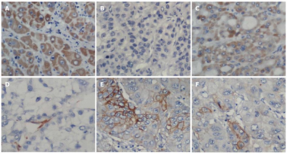
Figure 1 Representative images of hepatocellular carcinoma tissue.
Representative images of hepatocellular carcinoma (HCC) tissue showing positive and negative cytoplasmic staining for CD133 (A, B) and CD90 (C, D), or positive and negative membrane staining for epithelial cell adhesion molecule (EpCAM) (E, F). Magnification, × 400.
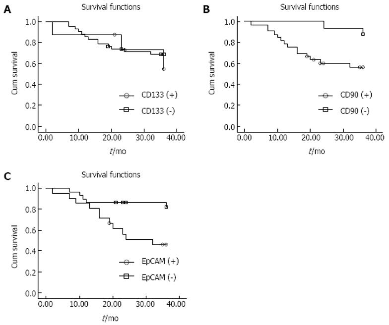
Figure 2 Overall survival curves.
A: For patients whose hepatocellular carcinoma (HCC) tissue was positive or negative for CD133 expression. The two curves were not significantly different, based on the log-rank test (P = 0.732); B: For patients whose HCC tissue was positive or negative for CD90 expression. Survival times were significantly shorter for CD90-positive patients (P = 0.018); C: For patients whose HCC tissue was positive or negative for epithelial cell adhesion molecule (EpCAM) expression. Survival times were significantly shorter for EpCAM-positive patients (P = 0.010).
- Citation: Guo Z, Li LQ, Jiang JH, Ou C, Zeng LX, Xiang BD. Cancer stem cell markers correlate with early recurrence and survival in hepatocellular carcinoma. World J Gastroenterol 2014; 20(8): 2098-2106
- URL: https://www.wjgnet.com/1007-9327/full/v20/i8/2098.htm
- DOI: https://dx.doi.org/10.3748/wjg.v20.i8.2098


