Copyright
©2014 Baishideng Publishing Group Co.
World J Gastroenterol. Feb 21, 2014; 20(7): 1822-1832
Published online Feb 21, 2014. doi: 10.3748/wjg.v20.i7.1822
Published online Feb 21, 2014. doi: 10.3748/wjg.v20.i7.1822
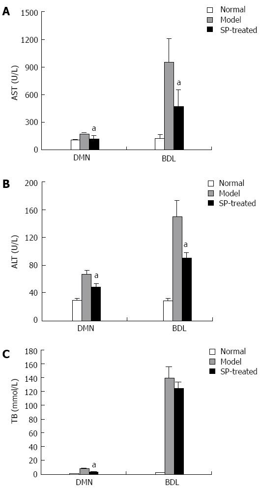
Figure 1 Sophocarpine ameliorates liver function in fibrotic rats.
Serum was collected from each group of rats. AST (A), ALT (B) and TB (C) levels were determined to assess liver function in the sophocarpine-treated group compared to each model group (aP < 0.05, by two-tailed Student’s t test). ALT: Alanine aminotransfer; AST: Aspartate aminotransferase; TB: Total bilirubin; DMN: Dimethylnitrosamine; BDL: Bile duct ligation.
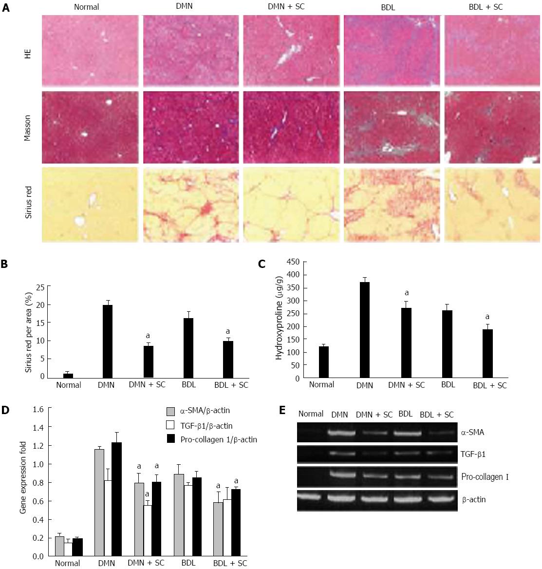
Figure 2 Sophocarpine attenuates hepatic fibrosis induced by dimethylnitrosamine or bile duct ligation in rats.
DMN and BDL were used to construct two types of hepatic fibrosis models to evaluate the therapeutic effect of sophocarpine. A: Liver fibrosis in each group was assessed by HE (× 40), Masson’s trichrome (× 40) and Sirius red staining (× 40); B: The percentage of Sirius-red in fibrotic livers was quantified using an image analysis system (aP < 0.05); C: The amount of hydroxyproline in fibrotic livers was detected in the sophocarpine-treated group compared with each model group (aP < 0.05); D, E: Real-time RT-PCR was employed to examine the expression of α-SMA, TGF-β and pro-collagen I in fibrotic livers following sophocarpine administration compared with the control models (aP < 0.05 by two-tailed Student’s t test). DMN: Dimethylnitrosamine; BDL: Bile duct ligation; TGF-β: Transforming growth factor-β; RT-PCR: Reverse transcription-polymerase chain reaction; SMA: Smooth muscle actin.
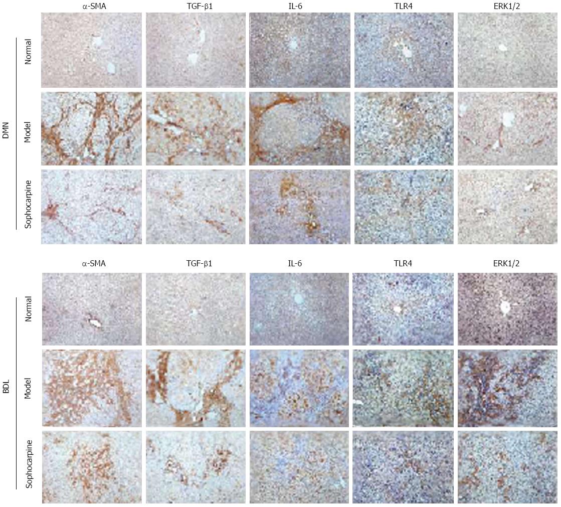
Figure 3 Expression of pro-fibrotic cytokines and toll-like receptor 4 signaling pathway related-proteins is suppressed in sophocarpine-treated rats.
Immunochemical analysis of the protein expression of α-SMA, TGF-β1, IL-6, TLR4 and ERK1/2 in the liver tissue of each group was performed as described in Materials and Methods. The results show the protein expression of α-SMA (× 200), TGF-β1 (× 200), IL-6 (× 200), TLR4 (× 200) and ERK1/2 (× 200) in the fibrotic livers of each group. TLR4: Toll-like receptor 4; IL-6: Interleukin-6; TGF-β: Transforming growth factor-β; SMA: Smooth muscle actin.
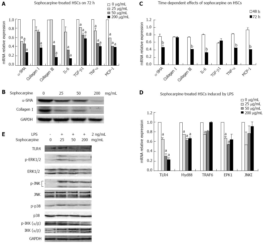
Figure 4 Sophocarpine inhibits the activation of hepatic stellate cells by blocking the lipopolysaccharide-induced toll-like receptor 4 signaling pathway.
Primary HSCs were isolated and plated in 6-well plates (1 × 10⁶ cells/well). Forty-eight hours later, the HSCs were treated with a gradient concentration of sophocarpine for 48 or 72 h. A: Real-time reverse transcription-polymerase chain reaction (RT-PCR) was performed to analyze the mRNA levels of α-SMA, collagen I, collagen III, IL-6, TGF-β1, TNF-α and MCP-1 in HSCs treated with a gradient concentration of sophocarpine for 72 h (compared to 0 μg/mL, aP < 0.05); B: Immunoblots of α-SMA, collagen I and GAPDH were detected by Western blot from HSCs treated with a gradient concentration of sophocarpine for 72 h; C: The mRNA expression of the above genes was detected in HSCs treated with sophocarpine (50 mg/mL) at 72 h compared to that at 48 h, bP < 0.05; D, E: Gradient concentration sophocarpine-treated HSCs were incubated with LPS (2 ng/mL), and real-time RT-PCR (compared to 0 μg/mL, aP < 0.05, D) and Western blot analysis (E) were employed to detect the expression of TLR4 pathway-related genes at the gene and protein levels (P value by two-tailed Student’s t test). LPS: Lipopolysaccharide; HSCs: Hepatic stellate cells; TGF-β: Transforming growth factor-β; IL-6: Interleukin-6; SMA: Smooth muscle actin; TNF: Tumor necrosis factor; MCP: Monocyte chemoattractant protein.
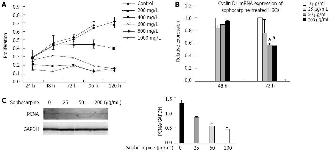
Figure 5 Sophocarpine suppresses the proliferation of hepatic stellate cells.
A: Activated HSCs were treated with a gradient concentration of sophocarpine and the proliferation of HSCs was assessed using the CCK-8 kit; B: Real-time polymerase chain reaction was performed to examine the expression of Cyclin D1 in HSCs after treatment with a gradient concentration of sophocarpine (aP < 0.05); C: Western blot was employed to detect PCNA expressed in HSCs after treatment with sophocarpine. HSCs: Hepatic stellate cells; PCNA: Proliferating cell nuclear antigen.
- Citation: Qian H, Shi J, Fan TT, Lv J, Chen SW, Song CY, Zheng ZW, Xie WF, Chen YX. Sophocarpine attenuates liver fibrosis by inhibiting the TLR4 signaling pathway in rats. World J Gastroenterol 2014; 20(7): 1822-1832
- URL: https://www.wjgnet.com/1007-9327/full/v20/i7/1822.htm
- DOI: https://dx.doi.org/10.3748/wjg.v20.i7.1822


