Copyright
©2014 Baishideng Publishing Group Co.
World J Gastroenterol. Feb 7, 2014; 20(5): 1361-1364
Published online Feb 7, 2014. doi: 10.3748/wjg.v20.i5.1361
Published online Feb 7, 2014. doi: 10.3748/wjg.v20.i5.1361
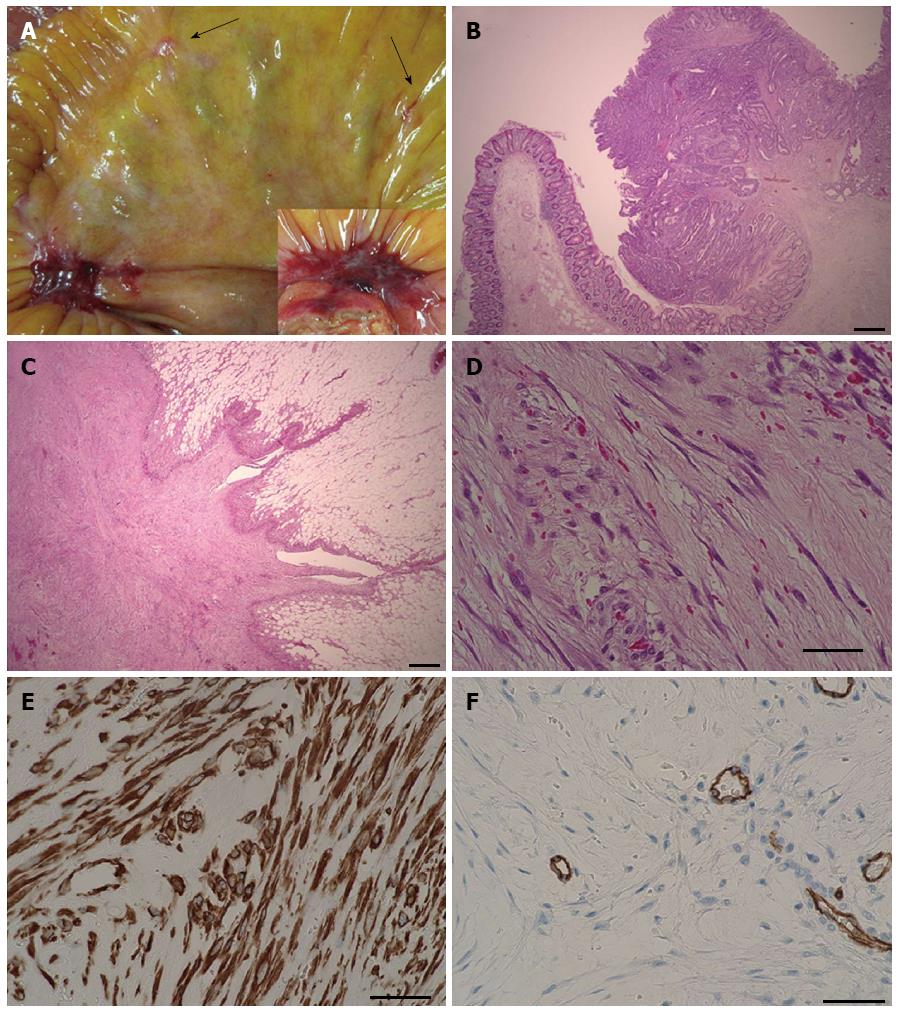
Figure 1 Intraoperative and pathological findings.
A: A relatively flat, reddish, and firm nodule was observed on the mesentery of the terminal ilium. The large nodule was accompanied by several whitish, smaller nodules (arrows). The inset shows another resected nodule; B: Resected type 1 ascending colon cancer infiltrating the submucosal layer; C: Lower magnification of nodular fasciitis showing growth in the adipose tissue, radiating outwardly in one part and growing expansively in another part. The margin of the nodule is not clear or encapsulated. Part of the nodule has sparse proliferation of fibroblast-like cells (less-pink area, center), while other parts have a quite dense striform proliferative pattern (pink-red color area); D: Higher magnification of nodular fasciitis demonstrating spindle-shape cells, which are arranged haphazardly throughout the abundant myxomatous interstitial ground substance. Numerous capillaries and extravasation of erythrocytes are visible. Some nuclei are plump, but do not have the hyperchromatic or bizarre pleomorphic features characteristically observed in sarcomas; E: The cytosol of the cells is positive for α-SMA staining, suggesting myofibroblastic differentiation; F: The nodule is negative for CD34, except for endothelial-cell CD34. The mucoid materials on the left side separate the thin spindle-shaped fibroblast-like cells, while the right side of the cells have plump nuclei. The bars for B and C are 500 μm, and the bars for D to F are 50 μm. α-SMA: α-smooth muscle actin.
- Citation: Shiga M, Okamoto K, Matsumoto M, Maeda H, Dabanaka K, Namikawa T, Uemura S, Munekage M, Kobayashi M, Hanazaki K. Nodular fasciitis in the mesentery, a differential diagnosis of peritoneal carcinomatosis. World J Gastroenterol 2014; 20(5): 1361-1364
- URL: https://www.wjgnet.com/1007-9327/full/v20/i5/1361.htm
- DOI: https://dx.doi.org/10.3748/wjg.v20.i5.1361


