Copyright
©2014 Baishideng Publishing Group Inc.
World J Gastroenterol. Nov 28, 2014; 20(44): 16779-16781
Published online Nov 28, 2014. doi: 10.3748/wjg.v20.i44.16779
Published online Nov 28, 2014. doi: 10.3748/wjg.v20.i44.16779
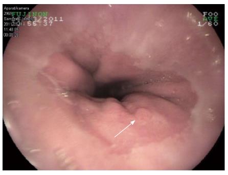
Figure 1 Endoscopic examination showing the location of the flat polypoid mass with benign features in the gastric cardia.
The lesion was approx. 10 mm below the “Z” line and measured approx. 7 mm in diameter.
Figure 2 Pancreatic heterotopia of the cardiac mucosa.
Most of the glandular structures visible below the foveolae correspond to pancreatic exocrine acini.
- Citation: Filip R, Walczak E, Huk J, Radzki RP, Bieńko M. Heterotopic pancreatic tissue in the gastric cardia: A case report and literature review. World J Gastroenterol 2014; 20(44): 16779-16781
- URL: https://www.wjgnet.com/1007-9327/full/v20/i44/16779.htm
- DOI: https://dx.doi.org/10.3748/wjg.v20.i44.16779


