Copyright
©2014 Baishideng Publishing Group Inc.
World J Gastroenterol. Nov 28, 2014; 20(44): 16665-16673
Published online Nov 28, 2014. doi: 10.3748/wjg.v20.i44.16665
Published online Nov 28, 2014. doi: 10.3748/wjg.v20.i44.16665
Figure 1 Estradiol and ER selective agonists impair cell proliferation in human LoVo cells.
A: Structures of 17β-estradiol, propylpyrazole-triol (PPT), a selective agonist of ERα, and diarylpropionitrile (DPN), a selective agonist of ERβ; B: LoVo cells cultured in DMEM were treated with 17β-estradiol, PPT and DPN for 24 h and 48 h and were then analyzed for cell viability by the MTT assay. aP < 0.05 vs control; bP < 0.001 vs control (means ± SE, n = 3).
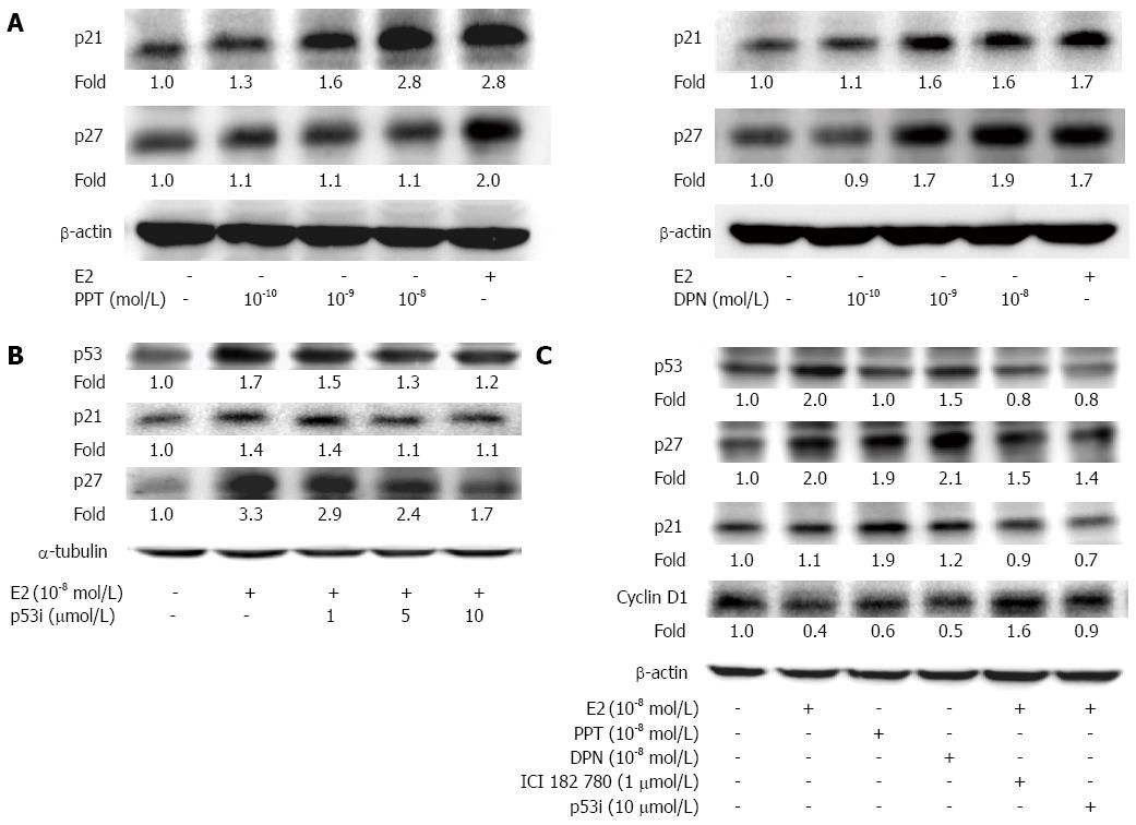
Figure 2 17β-estradiol and/or ER selective agonists upregulate p53 signaling proteins to change the cell cycle progression in human LoVo colorectal cancer cells.
A: LoVo cells were treated with E2 or various concentrations (10-10 mol/L, 10-9 mol/L and 10-8 mol/L) of PPT or DPN for 48 h; B: LoVo cells were pretreated with various concentrations (1 μmol/L, 5 μmol/L and 10 μmol/L) of a p53 inhibitor for 1 h followed by E2 (10-8 mol/L) treatment for 48 h; C: LoVo cells were pretreated with vehicle, the ER antagonist ICI 182780 (ICI), or a p53 inhibitor for 1h, followed by E2 administration for 48 h. Subsequently, cells were harvested and measured by Western blotting. A β-actin standard was used as a loading control for all proteins. All experiments were repeated twice with identical results.
Figure 3 17β-estradiol and ER selective agonists impair the Wnt-β-catenin signaling pathway in human LoVo colorectal cancer cells.
LoVo cells were treated with E2 (10-8 mol/L) or various concentrations (10-10 mol/L, 10-9 mol/L and 10-8 mol/L) of PPT (A) or DPN for 24 h (B). Cells were harvested and measured by Western blotting. An α-tubulin standard was used as a loading control for all proteins. All experiments were repeated twice with identical results.
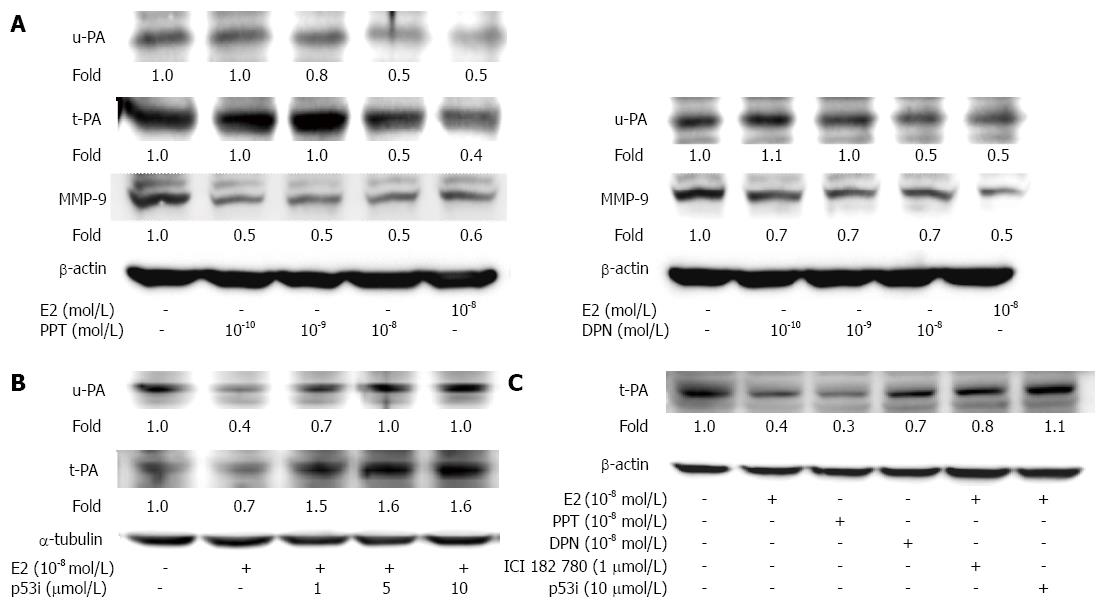
Figure 4 17β-estradiol and/or ER selective agonistsdown-regulate the protein levels of u-PA, t-PA and MMP-9 inLoVocells via p53.
A: LoVo cells were treated with E2 or various concentrations (10-10 mol/L, 10-9 mol/L and 10-8 mol/L) of PPT or DPN for 24 h; B: LoVo cells were pretreated with various concentrations (10-10 mol/L, 10-9 mol/L and 10-8 mol/L) of a p53 inhibitor for 1 h, followed by E2 administration for 24 h; C: LoVo cells were pretreated with vehicle, the ER antagonist ICI 182780, or a p53 inhibitor for 1 h, followed by E2 administration for 24 h. Cells were harvested and examined by Western blotting. A β-actin or α-tubulin standard was used as a loading control for all proteins. All experiments were repeated twice with identical results.
Figure 5 17β-estradiol inhibits MMP-2/9 activity in LoVo colorectal colon cells.
LoVo cells cultured in DMEM were treated with E2 (10-8 mol/L) for 24 h, and then, the culture medium was collected. Samples were electrophoresed without reduction on 8% SDS polyacrylamide gels copolymerized with 0.1% gelatin.
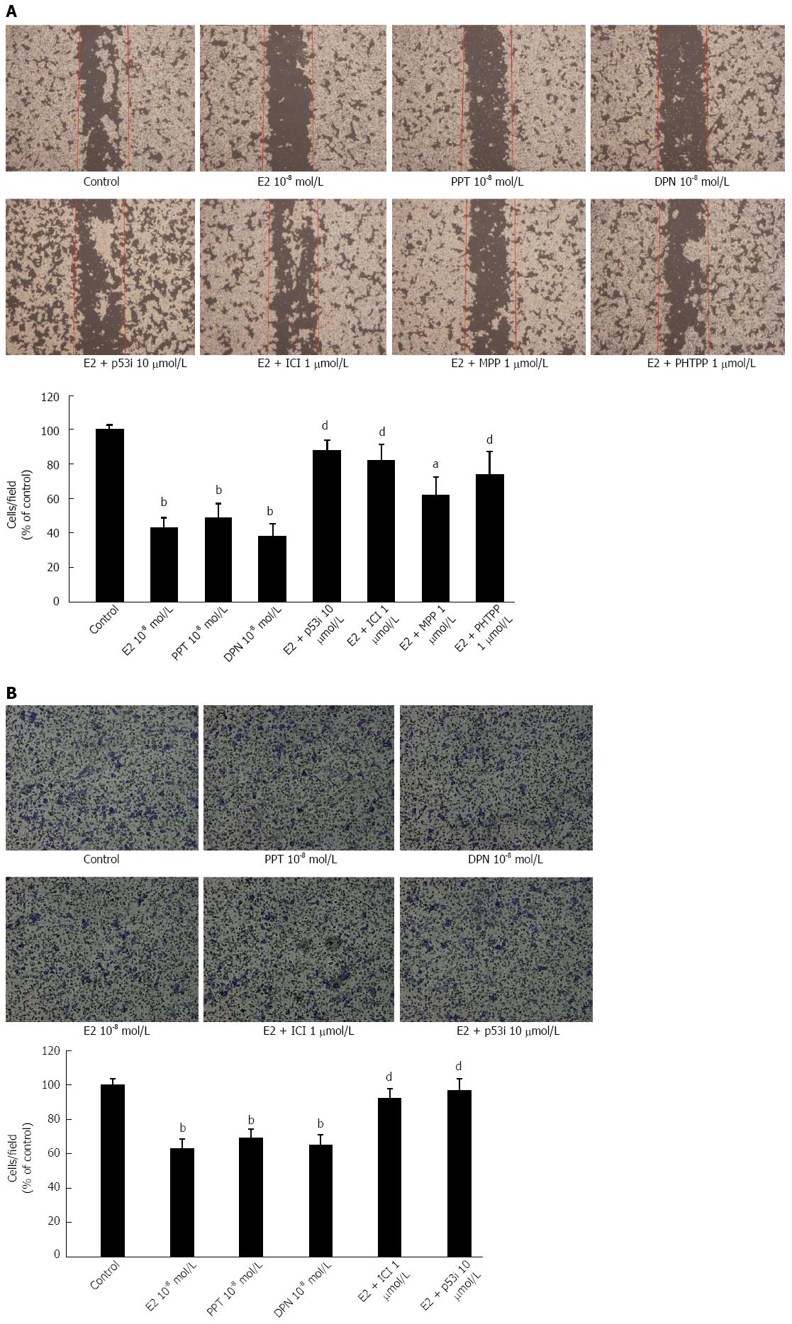
Figure 6 17β-estradiol and/or ER selective agonists inhibit human LoVo colorectal cancer cell motility through p53 signaling.
LoVo cells cultured in DMEM were pretreated with vehicle, ICI 182780 (ER antagonist 1 μmol/L), p53 inhibitor (10 μmol/L), MPP (ERα antagonist 1 μmol/L), or PHTPP (ERβ antagonist 1 μmol/L) for 1 h, followed by treatment with E2 (10-8 mol/L), PPT (10-8 mol/L) or DPN (10-8 mol/L) administration for 48h.The migration ability of LoVo cells was analyzed by a scratch motility assay (A) and a migration assay (B). The results were observed and analyzed with a fluorescence microscope. All experiments were repeated twice with identical results. aP < 0.05 vs control; bP < 0.01 vs control; dP < 0.01 vs E2.
- Citation: Hsu HH, Kuo WW, Ju DT, Yeh YL, Tu CC, Tsai YL, Shen CY, Chang SH, Chung LC, Huang CY. Estradiol agonists inhibit human LoVo colorectal-cancer cell proliferation and migration through p53. World J Gastroenterol 2014; 20(44): 16665-16673
- URL: https://www.wjgnet.com/1007-9327/full/v20/i44/16665.htm
- DOI: https://dx.doi.org/10.3748/wjg.v20.i44.16665


