Copyright
©2014 Baishideng Publishing Group Inc.
World J Gastroenterol. Oct 28, 2014; 20(40): 14717-14725
Published online Oct 28, 2014. doi: 10.3748/wjg.v20.i40.14717
Published online Oct 28, 2014. doi: 10.3748/wjg.v20.i40.14717
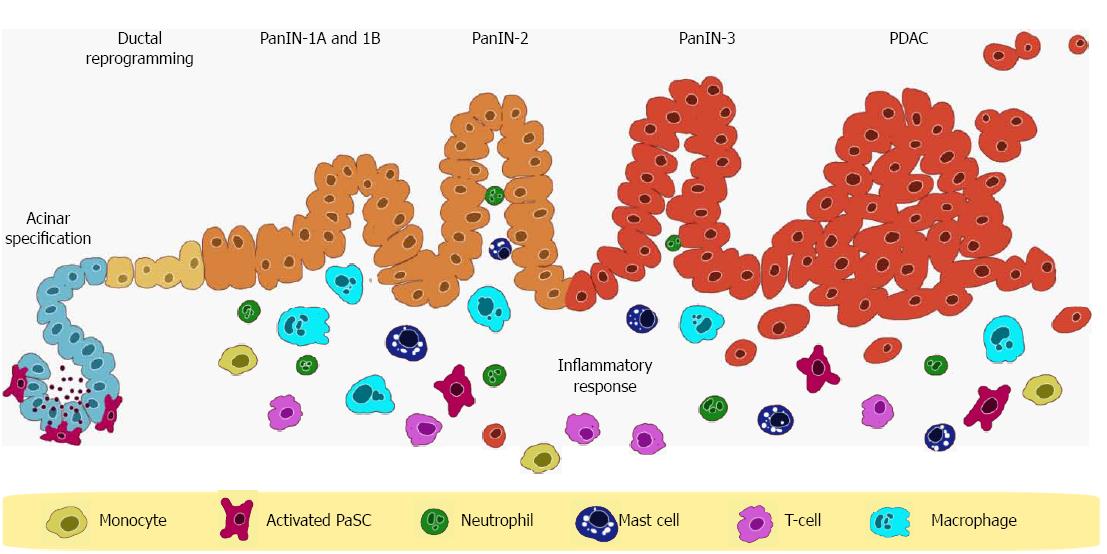
Figure 1 Example of pancreatic intraepithelial neoplasia lesion development to pancreatic ductal adenocarcinoma.
Inflammatory stimuli trigger the activation of pancreatic stellate cells (PaSC) surrounding acinus cells. Inflammatory cells (monocytes, T-cells, neutrophils, mast cells, and macrophages) gather in response and release ligands (interleukin-6) that activate STAT3 gene to promote pancreatic intraepithelial neoplasia (PanIN) development in susceptible tissue with oncogenic mutations such as the Kras. PDAC: Pancreatic ductal adenocarcinoma. Figure redrawn with permissions from Elsevier: [10] and Macmillan Publishers Ltd: [12].
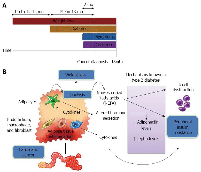
Figure 2 Symptoms of paraneoplastic type 3c diabetes preceding pancreatic cancer.
A: A comparison of weight-loss timeline to cancer-specific symptoms; B: Schematic representation of the cause for progressive weight reduction and insulin resistance. Adipose tissue inflammation triggers an alteration of adipocyte secretion and propagates pathogenic processes similar to type 2 diabetes, eventually leading to cachexia. Figures redrawn with permission from Macmillan Publishers Ltd: [17].
- Citation: Loc WS, Smith JP, Matters G, Kester M, Adair JH. Novel strategies for managing pancreatic cancer. World J Gastroenterol 2014; 20(40): 14717-14725
- URL: https://www.wjgnet.com/1007-9327/full/v20/i40/14717.htm
- DOI: https://dx.doi.org/10.3748/wjg.v20.i40.14717


