Copyright
©2014 Baishideng Publishing Group Inc.
World J Gastroenterol. Oct 21, 2014; 20(39): 14230-14236
Published online Oct 21, 2014. doi: 10.3748/wjg.v20.i39.14230
Published online Oct 21, 2014. doi: 10.3748/wjg.v20.i39.14230
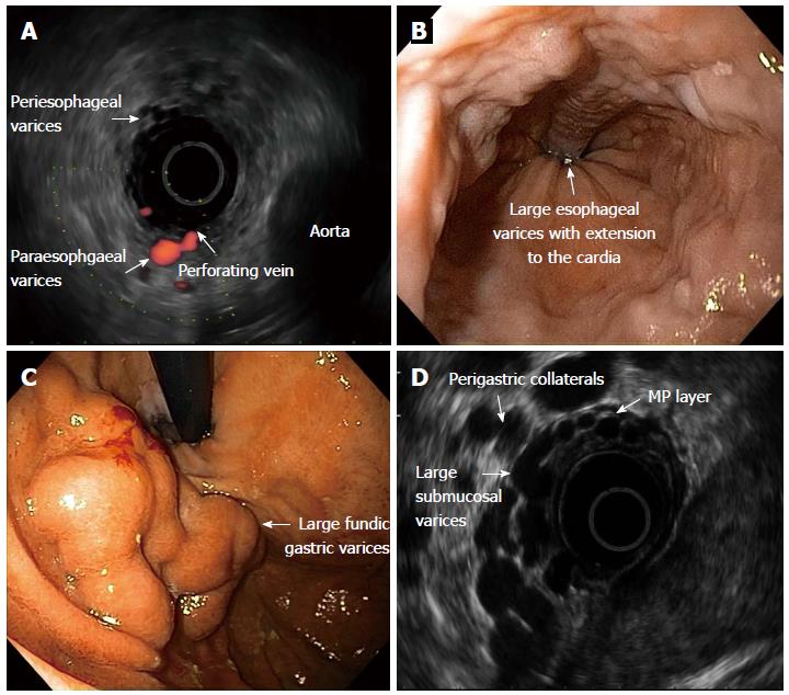
Figure 1 Anatomy of the distal esophagus and proximal stomach in portal hypertension.
A: Radial echoendoscope shows esophageal varices, periesophageal varices and paraesophgaeal varices with associated perforating vein; B: Upper endoscopy: Large esophageal varices extending to the cardia; C: Upper endoscopy: Type-2 large gastroesophageal varices; D: Radial echoendoscope shows large gastric and perigastric collaterals.
- Citation: Hammoud GM, Ibdah JA. Utility of endoscopic ultrasound in patients with portal hypertension. World J Gastroenterol 2014; 20(39): 14230-14236
- URL: https://www.wjgnet.com/1007-9327/full/v20/i39/14230.htm
- DOI: https://dx.doi.org/10.3748/wjg.v20.i39.14230


