Copyright
©2014 Baishideng Publishing Group Inc.
World J Gastroenterol. Oct 7, 2014; 20(37): 13607-13614
Published online Oct 7, 2014. doi: 10.3748/wjg.v20.i37.13607
Published online Oct 7, 2014. doi: 10.3748/wjg.v20.i37.13607
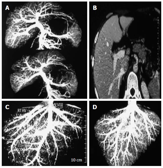
Figure 1 Examples of excluded donors due to unsuitable anatomical variations as shown by imaging studies.
(A) Computerized tomographic (CT) portography showing the right portal vein arises from the left portal vein (B) CT volumetry showing that drainage of right lobe is mainly through the middle hepatic vein (C) CT venography showing drainage of the right lobe through multiple hepatic veins (D) CT venography showing small right hepatic vein and inferior right hepatic vein draining into the middle hepatic vein.
- Citation: Wahab MA, Hamed H, Salah T, Elsarraf W, Elshobary M, Sultan AM, Shehta A, Fathy O, Ezzat H, Yassen A, Elmorshedi M, Elsaadany M, Shiha U. Problem of living liver donation in the absence of deceased liver transplantation program: Mansoura experience. World J Gastroenterol 2014; 20(37): 13607-13614
- URL: https://www.wjgnet.com/1007-9327/full/v20/i37/13607.htm
- DOI: https://dx.doi.org/10.3748/wjg.v20.i37.13607


