Copyright
©2014 Baishideng Publishing Group Inc.
World J Gastroenterol. Sep 14, 2014; 20(34): 12359-12362
Published online Sep 14, 2014. doi: 10.3748/wjg.v20.i34.12359
Published online Sep 14, 2014. doi: 10.3748/wjg.v20.i34.12359
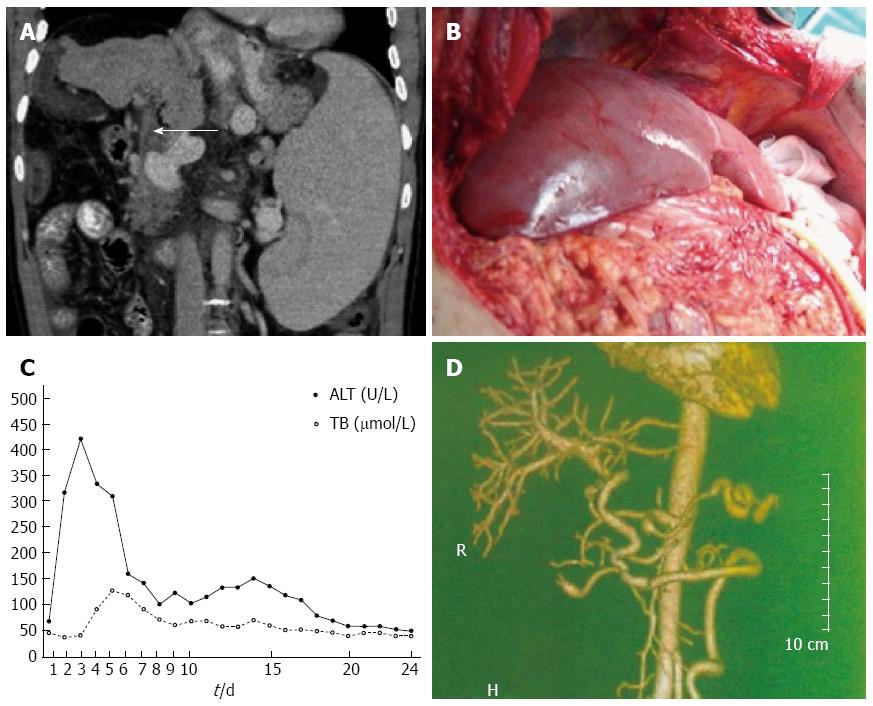
Figure 1 Case 1 imaging diagnosis.
A: Computed tomography scan of the upper abdomen (portal vein thrombi) (arrow); B: Donor liver after the arterial venous circulation was opened; C: Alanine aminotransferase (ALT) and total bilirubin (TB) levels after transplantation; D: Computed tomography angiography of the upper abdomen (pedestrian vehicle accident anastomotic stoma).
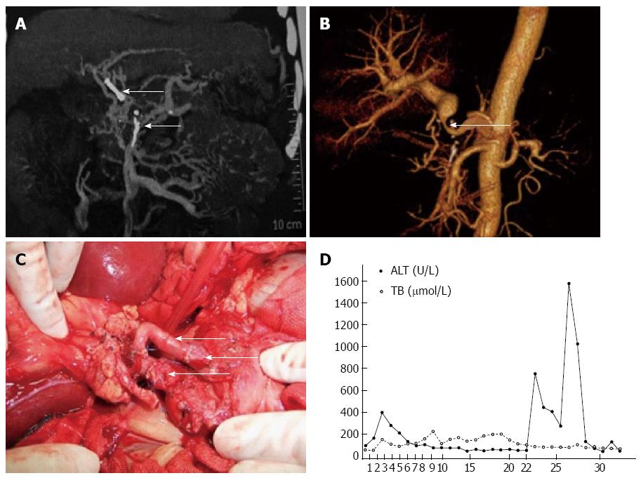
Figure 2 Case 2 imaging diagnosis.
A: Computed tomography scan of the upper abdomen (portal vein thrombi); B: Computed tomography angiography of the upper abdomen [pedestrian vehicle accident (PVA) anastomotic stoma]; C: PVA anastomotic stoma (liver artery of the donor, gastroduodenal artery of the recipient and the common hepatic artery of the recipient); D: Alanine aminotransferase (ALT) and total bilirubin (TB) levels after transplantation.
- Citation: Zhang K, Jiang Y, Lv LZ, Cai QC, Yang F, Hu HZ, Zhang XJ. Portal vein arterialization technique for liver transplantation patients. World J Gastroenterol 2014; 20(34): 12359-12362
- URL: https://www.wjgnet.com/1007-9327/full/v20/i34/12359.htm
- DOI: https://dx.doi.org/10.3748/wjg.v20.i34.12359


