Copyright
©2014 Baishideng Publishing Group Inc.
World J Gastroenterol. Sep 7, 2014; 20(33): 11650-11670
Published online Sep 7, 2014. doi: 10.3748/wjg.v20.i33.11650
Published online Sep 7, 2014. doi: 10.3748/wjg.v20.i33.11650
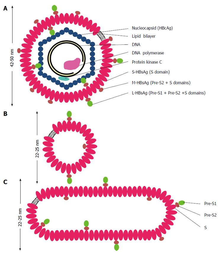
Figure 1 Schematic representations of hepatitis B virus virion (A), spherical (B) and filamentous (C) particles.
The envelope of hepatitis B virus (HBV) virion or the Dane particle (A) contains three forms of hepatitis B surface antigen (HBsAg): large (L-) HBsAg has the PreS1, PreS2 and S domains; middle (M-) HBsAg contains the PreS2 and S domains; and small (S-) HBsAg has only the S domain. The representations of the L-, M-, and S-HBsAg have no quantitative or positional significance. The L-HBsAg interacts with the viral capsid which is made of many copies of the core protein (HBcAg). The capsid encapsidates a partially double stranded DNA molecule, a DNA polymerase containing the primase and reverse transcriptase activities. The protein kinase C phosphorylates the capsid protein. The diameter of HBV virion is about 42 nm when it is stained negatively and observed under a transmission electron microscope, but it appears bigger in cryo-electron microscopy. The spherical (B) and filamentous (C) particles have a diameter about 22 nm in negative staining and appear bigger in cryo-electron microscopy. The length of the filamentous particle varies. Both the non-infectious particles contain the L-, M-, S-HBsAg and lipid.
Figure 2 Envelope proteins of hepatitis B virus.
The translation products of the HBsAg gene are shown as lines of different thickness. Small (S-), middle (M-) and large (L-) HBsAg are translated from a common open reading frame of the HBsAg gene by the use of three in-frame initiation codons (1) at the N-termini of PreS1, PreS2 and S. G: Glycosylation; MYR: Myristic acid; HBV: Hepatitis B virus; HBsAg: Hepatitis B surface antigen.
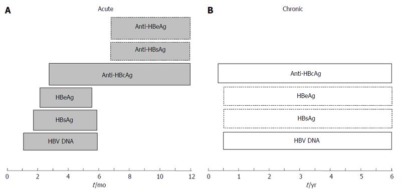
Figure 3 Hepatitis B virus markers in sera during acute (A) and chronic (B) infections.
The periods in months (acute infection) and years (chronic infection) are indicated below the boxes containing the markers. The presence of the markers indicated in boxes with broken lines varies in individuals. HBV: Hepatitis B virus; HBsAg: Hepatitis B surface antigen; HBeAg: Hepatitis B e antigen; HBcAg: Hepatitis B core antigen.
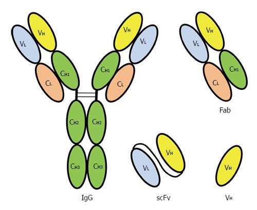
Figure 4 Recombinant antibodies displayed on bacteriophage compared with an immunoglobulin G molecule.
Chain structure of a human immunoglobulin G (IgG) molecule. V: Variable region; C: Constant region; H: Heavy chain; L: Light chain; VH: Variable region of heavy chain; Fab: Fragment antigen binding; scFv: Single chain fragment variable, the VL and VH are linked by a peptide linker.
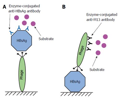
Figure 5 Phage-ELISA for detecting hepatitis B virus surface antigen.
A: Major steps used in an assay for detecting hepatitis B virus surface antigen (HBsAg) by immobilizing phage carrying the peptide on a solid support. The phage harboring the peptide is immobilized on a solid support. The unsaturated area of the solid phase is blocked by 10% milk diluent. The sample containing HBsAg is added to interact with the peptide. Mouse anti-HBsAg antibody is added to interact with the captured HBsAg. Then anti-mouse antibody conjugated to an enzyme is added to interact with the anti-HBsAg antibody. Finally, substrates for the enzyme are added to produce a measurable signal; B: Major steps used in an assay for detecting HBsAg immobilized on a solid support using a phage carrying the peptide. A sample containing HBsAg is immobilized on a solid support. The unsaturated area of the solid phase is blocked by 10% milk diluent. The phage harboring the peptide is added to interact with immobilized HBsAg. Anti-M13 phage antibody conjugated to an enzyme is added to interact with the bound phage and substrates for the enzyme are added to produce a measurable signal. ELISA: Enzyme-Linked Immunosorbent Assay.
- Citation: Tan WS, Ho KL. Phage display creates innovative applications to combat hepatitis B virus. World J Gastroenterol 2014; 20(33): 11650-11670
- URL: https://www.wjgnet.com/1007-9327/full/v20/i33/11650.htm
- DOI: https://dx.doi.org/10.3748/wjg.v20.i33.11650


