Copyright
©2014 Baishideng Publishing Group Inc.
World J Gastroenterol. Aug 14, 2014; 20(30): 10470-10477
Published online Aug 14, 2014. doi: 10.3748/wjg.v20.i30.10470
Published online Aug 14, 2014. doi: 10.3748/wjg.v20.i30.10470
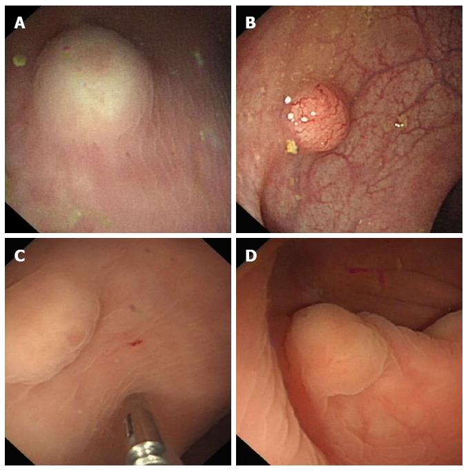
Figure 1 Endoscopic characteristics of rectal neuroendocrine neoplasms.
A: A yellowish hemispherical bulge overlaying intact mucosa; B: The appearance of a pyknic vascular net on the surface of a hemispherical bulge; C: A hummocky bulge with erosion on the surface; D: A flat-shaped neuroendocrine neoplasm.
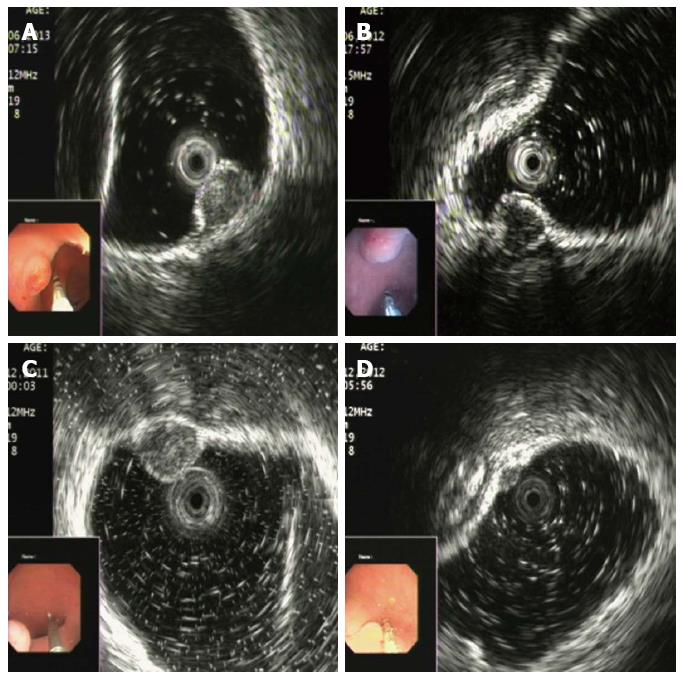
Figure 2 Endoscopic ultrasonography findings of rectal neuroendocrine neoplasms.
A: An egg-shaped lesion within the mucosa with an intermediate echo pattern and distinct border; B: A homogenous hypoechoic lesion with a distinct border within the submucosa; C: A round medium-echo lesion with a distinct border within the submucosa; D: A nodular neuroendocrine neoplasm located in both the mucosa and submucosa.
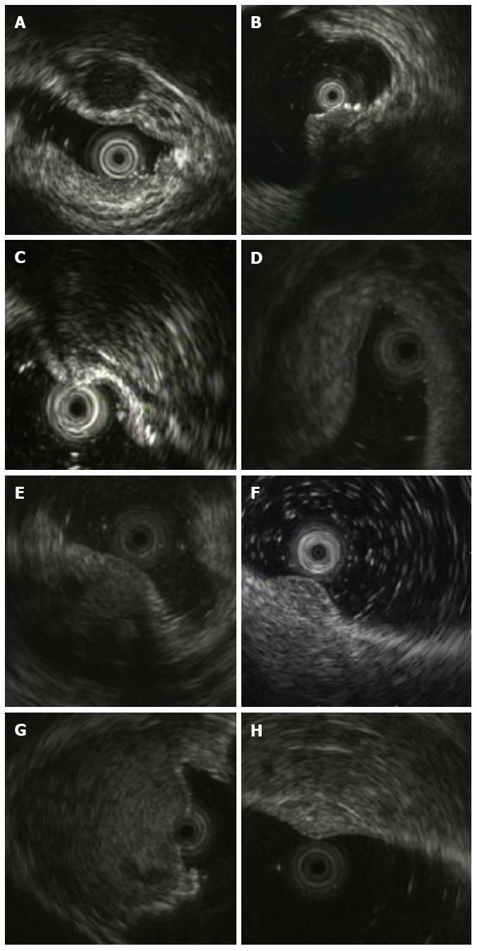
Figure 3 Endoscopic ultrasonography characteristics of 8 rectal non-neuroendocrine neoplasm subepithelial lesions misdiagnosed as neuroendocrine neoplasms.
A: GIST confirmed by pathology after endoscopic submucosal dissection (ESD) presents as a round homogenous hypoechoic lesion within the submucosa; B: Rectal endometriosis identified by endoscopic biopsy shows a hypoechoic lesion with full-thickness infiltration, and irregular and undefined margins, extending outside the rectal wall; C: A nodule with fibrosis and degeneration shown as a heterogeneous hyperechoic nodule within the submucosa with a blurry border on EUS; D: EUS characteristics of an inflammatory lesion identified by pathology from excisional specimen; E: A metastatic tumor secondary to porta carcinoma of the bile duct mimicking NEN with local infiltration of the rectal wall; F: Rectal lymphoma shown as a heterogeneous hypoechoic lesion infiltrating the 2nd-3rd-4th wall layers; G: Rectal neurilemmoma shown as a heterogeneous intermediate lesion with an irregular anechoic area representing necrosis; H: Rectal hemangioma shown as an SEL with honeycomb echo denoting sinusoidal blood. EUS: Endoscopic ultrasonography; SEL: Subepithelial lesion; NEN: Neuroendocrine neoplasm; GIST: Gastric small gastrointestinal tumor.
- Citation: Chen HT, Xu GQ, Teng XD, Chen YP, Chen LH, Li YM. Diagnostic accuracy of endoscopic ultrasonography for rectal neuroendocrine neoplasms. World J Gastroenterol 2014; 20(30): 10470-10477
- URL: https://www.wjgnet.com/1007-9327/full/v20/i30/10470.htm
- DOI: https://dx.doi.org/10.3748/wjg.v20.i30.10470


