Copyright
©2014 Baishideng Publishing Group Inc.
World J Gastroenterol. Jul 21, 2014; 20(27): 9200-9204
Published online Jul 21, 2014. doi: 10.3748/wjg.v20.i27.9200
Published online Jul 21, 2014. doi: 10.3748/wjg.v20.i27.9200
Figure 1 Abdominal imaging and endoscopic ultrasound of a patient 1 undergoing routine surveillance for a pancreatic cyst.
(A) and (B) demonstrate the cystic lesion in the head/uncinate process of the pancreas as seen on computed tomography scan (arrow); and seen on endoscopic ultrasound (C). Image (D) shows a mass1 within the head of the pancreas which was separate to the cyst.
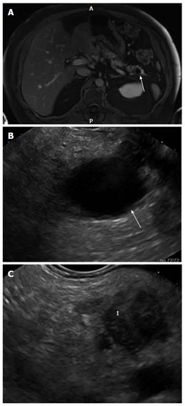
Figure 2 Abdominal imaging and endoscopic ultrasound of the pancreatic remnant in patient 2 who was status post pancreaticoduodenctomy for intraductal papillary mucinous neoplasm.
A: Magnetic resonance imaging of the remnant pancreas performed 6 mo before the endoscopic ultrasound (EUS). The arrow demonstrates the cyst in the tail of the pancreas; B: EUS image of the cyst in the tail of the pancreas (arrow) corresponding to the MRI image seen in (A); C: Demonstrates an ill-defined hypoechoic area1 in the body of the pancreas.
- Citation: Law JK, Wolfgang CL, Weiss MJ, Lennon AM. Concomitant pancreatic adenocarcinoma in a patient with branch-duct intraductal papillary mucinous neoplasm. World J Gastroenterol 2014; 20(27): 9200-9204
- URL: https://www.wjgnet.com/1007-9327/full/v20/i27/9200.htm
- DOI: https://dx.doi.org/10.3748/wjg.v20.i27.9200


