Copyright
©2014 Baishideng Publishing Group Inc.
World J Gastroenterol. Jul 21, 2014; 20(27): 9026-9037
Published online Jul 21, 2014. doi: 10.3748/wjg.v20.i27.9026
Published online Jul 21, 2014. doi: 10.3748/wjg.v20.i27.9026
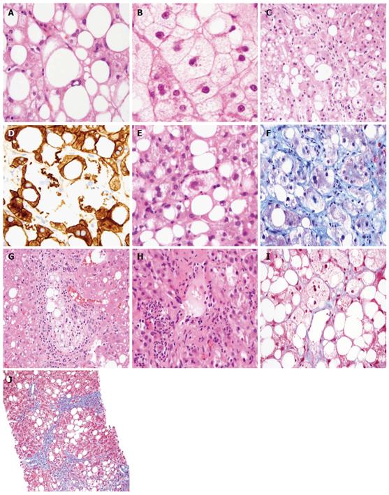
Figure 1 Histologic features, grading, and staging of nonalcoholic fatty liver disease.
A: Mixed large and small droplet steatosis, single droplet, with nucleus pushed to one side, HE stain, 600 ×; B: Microvesicular steatosis, nuclei in the center with foamy cytoplasm, and megamitochondria HE stain, 600 ×; C: Ballooned hepatocytes with flocculent cytoplasm, HE stain, 600 ×; D: Loss of cytoplasmic expression of keratin 8/18 in ballooned hepatocytes, 600 ×; E: Mallory-Denk body, HE stain, 600 ×; F: Mallory-Denk body in blue-green color and dense perisinusoidal fibrosis, Trichrome stain, 600 ×; G: Portal lipogranuloma, HE stain, 400 ×; H: Mallory-Denk bodies and satellitosis HE stain, 600 ×; I: Delicate perisinusoidal fibrosis, Trichrome stain, 600 ×; J: Bridging fibrosis, Trichrome stain, 200 ×.
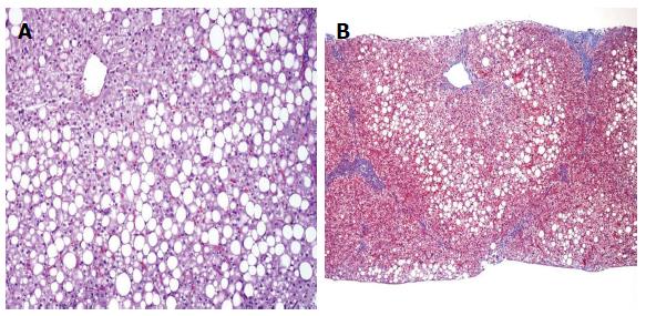
Figure 2 Pediatric nonalcoholic fatty liver disease.
A: Periportal accentuation of steatosis with sparing of zone 3, pediatric nonalcoholic fatty liver disease (NAFLD), HE stain, 200 ×; B: Portal fibrosis without zone 3 perisinusoidal fibrosis, pediatric NAFLD, Trichrome stain, 100 ×.
- Citation: Nalbantoglu I, Brunt EM. Role of liver biopsy in nonalcoholic fatty liver disease. World J Gastroenterol 2014; 20(27): 9026-9037
- URL: https://www.wjgnet.com/1007-9327/full/v20/i27/9026.htm
- DOI: https://dx.doi.org/10.3748/wjg.v20.i27.9026


