Copyright
©2014 Baishideng Publishing Group Inc.
World J Gastroenterol. Jul 7, 2014; 20(25): 8309-8311
Published online Jul 7, 2014. doi: 10.3748/wjg.v20.i25.8309
Published online Jul 7, 2014. doi: 10.3748/wjg.v20.i25.8309
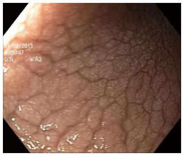
Figure 1 Endoscopic appearance of the stomach body.
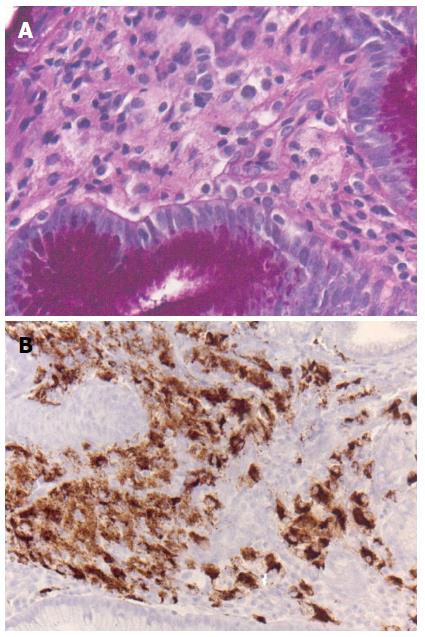
Figure 2 Histological findings in gastric biopsy.
A: Lamina propria infiltrated by foamy histiocytes negative for PAS (PAS stain, × 200); B: Foamy cells stained positive for CD 68 (CD68 stain, × 100).
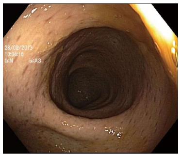
Figure 3 Colonoscopy showing numerous small round hyper-pigmented lesions.
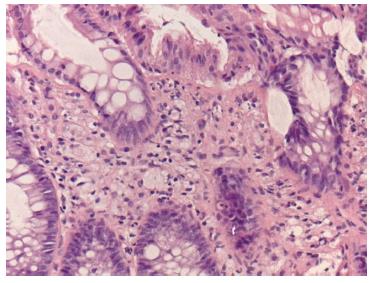
Figure 4 Histology of colonic mucosa showing histiocytic infiltration of lamina propria (HE stain, × 100).
- Citation: Ben-yaakov G, Munteanu D, Sztarkier I, Fich A, Schwartz D. Erdheim chester - A rare disease with unique endoscopic features. World J Gastroenterol 2014; 20(25): 8309-8311
- URL: https://www.wjgnet.com/1007-9327/full/v20/i25/8309.htm
- DOI: https://dx.doi.org/10.3748/wjg.v20.i25.8309


