Copyright
©2014 Baishideng Publishing Group Co.
World J Gastroenterol. Jan 14, 2014; 20(2): 603-606
Published online Jan 14, 2014. doi: 10.3748/wjg.v20.i2.603
Published online Jan 14, 2014. doi: 10.3748/wjg.v20.i2.603
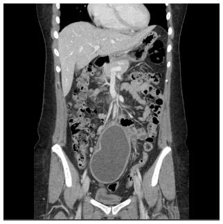
Figure 1 A contrast-enhanced coronal image of computed tomography reveals a well-circumscribed and poorly attenuated dumbbell-shaped mass exhibiting peripheral enhancement and a trilaminar appearance in the pelvic cavity.
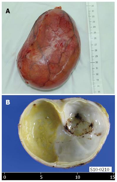
Figure 2 Gross findings of the resected mass.
The 12-cm-long cystic mass has a smooth pinkish outer surface (A), and the cavity is uniloculated and incompletely septated (B).
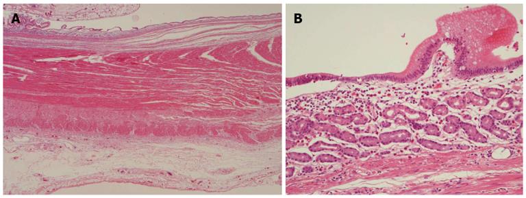
Figure 3 Microphotographs of the cystic wall.
The wall consists of mucosa, submucosa, muscle layers, and serosa (A, HE stain, × 10), with regions of gastric-type mucosa containing oxyntic glands (B, HE stain, × 100).
- Citation: Park JY, Her KH, Kim BS, Maeng YH. A completely isolated intestinal duplication cyst mimicking ovarian cyst torsion in an adult. World J Gastroenterol 2014; 20(2): 603-606
- URL: https://www.wjgnet.com/1007-9327/full/v20/i2/603.htm
- DOI: https://dx.doi.org/10.3748/wjg.v20.i2.603


