Copyright
©2014 Baishideng Publishing Group Inc.
World J Gastroenterol. May 21, 2014; 20(19): 5930-5934
Published online May 21, 2014. doi: 10.3748/wjg.v20.i19.5930
Published online May 21, 2014. doi: 10.3748/wjg.v20.i19.5930
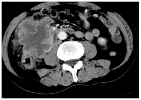
Figure 1 Abdominal computed tomography which revealed a large soft tissue mass of 7.
6 cm × 8.5 cm in size in the ascending colon.
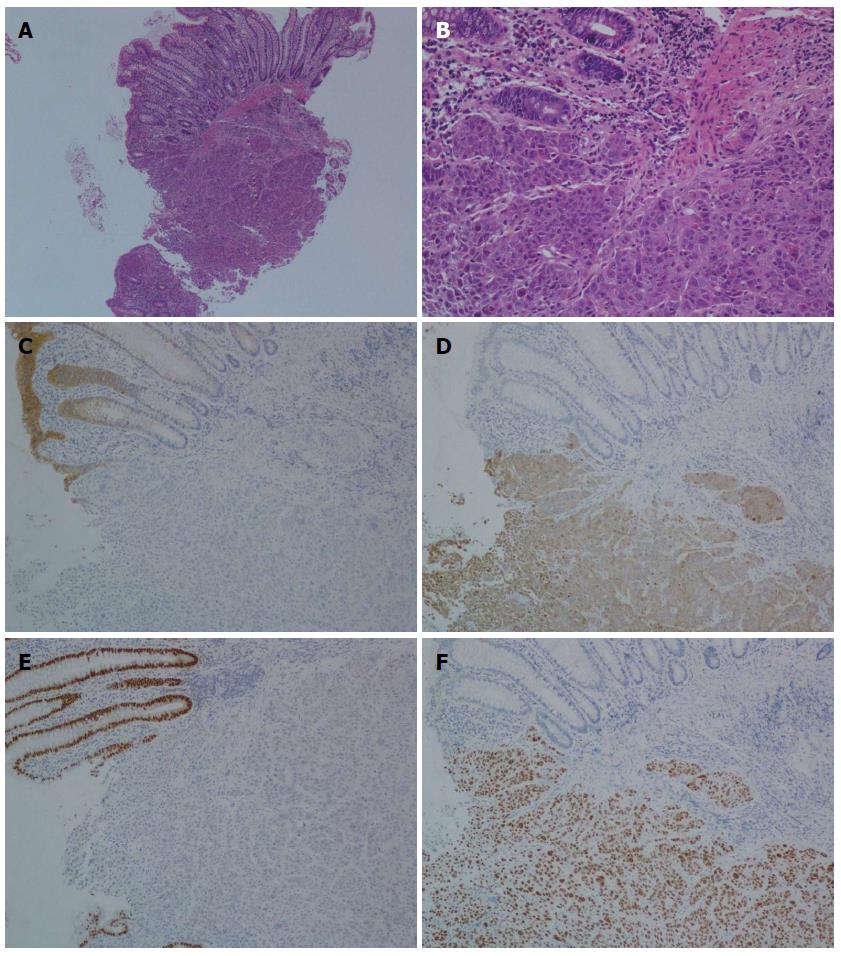
Figure 2 Histological findings.
A: Poorly-differentiated squamous cell carcinoma metastatic to the descending colon [hematoxylin eosin (HE) × 40]; B: Cancercells infiltrating mucosa of the descending colon (HE × 200); C: Negative immunohistochemical staining for CK 20 (× 100); D: Positive immunohistochemical staining for CK 5/6 (× 100); E: Negative immunohistochemical staining for CDX-2 (× 100); F: Positive immunohistochemical staining for p63 (× 100).
- Citation: Lou HZ, Wang CH, Pan HM, Pan Q, Wang J. Colonic metastasis after resection of primary squamous cell carcinoma of the lung: A case report and literature review. World J Gastroenterol 2014; 20(19): 5930-5934
- URL: https://www.wjgnet.com/1007-9327/full/v20/i19/5930.htm
- DOI: https://dx.doi.org/10.3748/wjg.v20.i19.5930


