Copyright
©2014 Baishideng Publishing Group Inc.
World J Gastroenterol. May 21, 2014; 20(19): 5924-5929
Published online May 21, 2014. doi: 10.3748/wjg.v20.i19.5924
Published online May 21, 2014. doi: 10.3748/wjg.v20.i19.5924
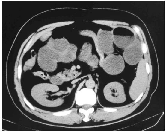
Figure 1 Abdominal computed tomography showed dilated loops of small bowel with multiple air fluids levels and collapsed distal small bowel, consistent with small bowel obstruction.
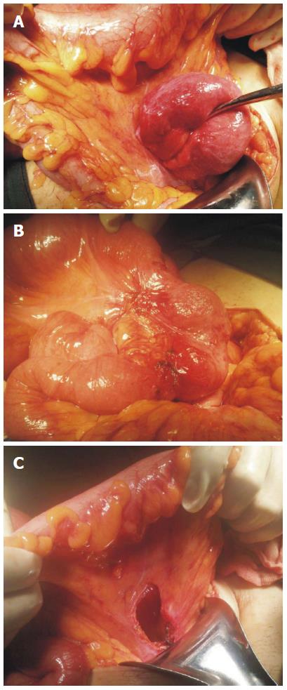
Figure 2 Sigmoid mesocolic hernia during laparotomy, with 15-cm loop of ileum herniated into the hiatus of the sigmoid mesocolon (A), herniated loop showing no gangrene, but the presence of a constriction ring (B), orifice about 2.
5 cm in diameter (C).
- Citation: Li B, Assaf A, Gong YG, Feng LZ, Zheng XY, Wu CN. Transmesosigmoid hernia: Case report and review of literature. World J Gastroenterol 2014; 20(19): 5924-5929
- URL: https://www.wjgnet.com/1007-9327/full/v20/i19/5924.htm
- DOI: https://dx.doi.org/10.3748/wjg.v20.i19.5924


