Copyright
©2014 Baishideng Publishing Group Inc.
World J Gastroenterol. May 21, 2014; 20(19): 5575-5582
Published online May 21, 2014. doi: 10.3748/wjg.v20.i19.5575
Published online May 21, 2014. doi: 10.3748/wjg.v20.i19.5575
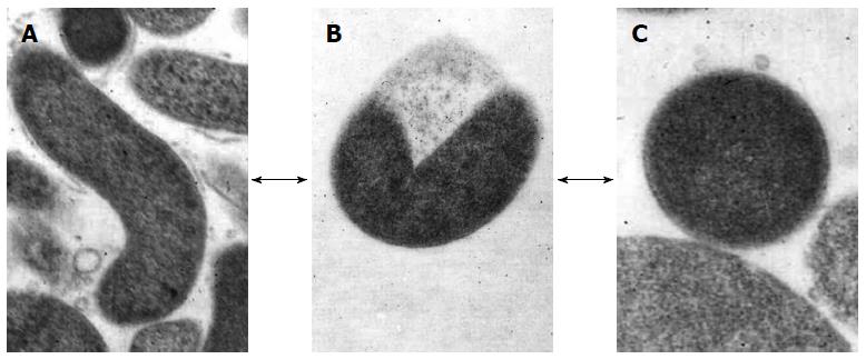
Figure 1 Morphological appearance of Helicobacter pylori.
A: Rod-shaped; B: U-shaped; C: Coccoid form. Arrows show the hypothetical alternative pathway between the different forms. Transmission electron microscope: original magnification × 20000.
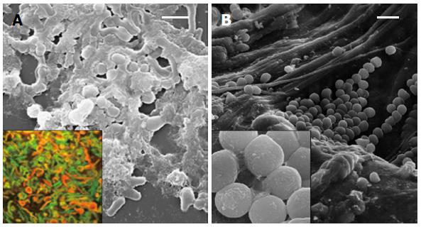
Figure 2 Biofilm of Helicobacter pylori.
A: Scanning electron micrograph of mature biofilm on a polystyrene surface with rod shaped and coccoid cells embedded in an abundant matrix. Insert: Confocal laser scanning microscopy image of mature in vitro biofilm with viable (green) and dead (red) cells, live/dead staining; B: Clusters of coccoid Helicobacter pylori cells in the gastric mucosa also embedded in a matrix (insert). Bars represent 5 μm.
-
Citation: Cellini L.
Helicobacter pylori : A chameleon-like approach to life. World J Gastroenterol 2014; 20(19): 5575-5582 - URL: https://www.wjgnet.com/1007-9327/full/v20/i19/5575.htm
- DOI: https://dx.doi.org/10.3748/wjg.v20.i19.5575


