Copyright
©2014 Baishideng Publishing Group Co.
World J Gastroenterol. Apr 28, 2014; 20(16): 4771-4777
Published online Apr 28, 2014. doi: 10.3748/wjg.v20.i16.4771
Published online Apr 28, 2014. doi: 10.3748/wjg.v20.i16.4771
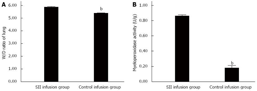
Figure 1 Change in lung wet-to-dry weight ratio and myeloperoxidase level.
Severe intraperitoneal infection (SII) lymph infusion increased inflammatory injury. Myeloperoxidase (MPO) activity (A) and wet-to-dry weight (W/D) ratio (B) were measured in lung collected from SII and control lymph-infused rats 4 h after infusion. The results (mean ± SD) are the summary of two separate experiments (n = 10 rats in each group). bP < 0.01 vs SII infusion group.
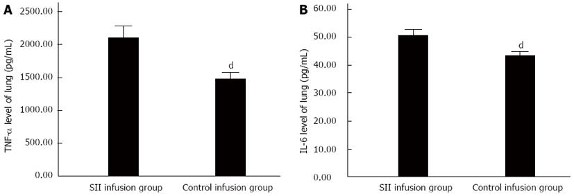
Figure 2 Tumor necrosis factor-α and interleukin-6 levels in lung tissues.
Severe intraperitoneal infection (SII) lymph infusion enhances sepsis-induced inflammatory cytokine responses in the lung. Concentrations of tumor necrosis factor-α (A) and interleukin (IL)-6 (B) were measured from lung extracts of SII and control lymph-infused rats. Data are mean ± SD of 10 rats in each group. Results are representative of two separate experiments. dP < 0.01 vs SII infusion group.
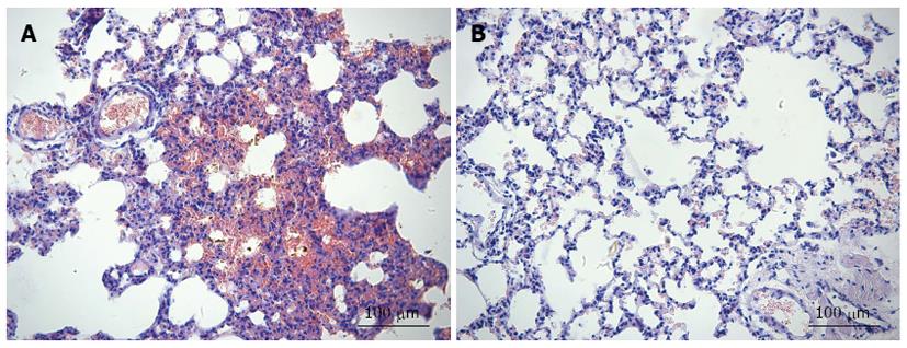
Figure 3 Tissue sections of lungs of severe intraperitoneal infection and control infusion rats stained with hematoxylin and eosin.
Evidence of increased interstitial edema and inflammatory cell infiltration was found in rats injected with SII lymph (A). In contrast, control lymph-infused rats showed no evidence of lung injury (B). Magnification, × 200.
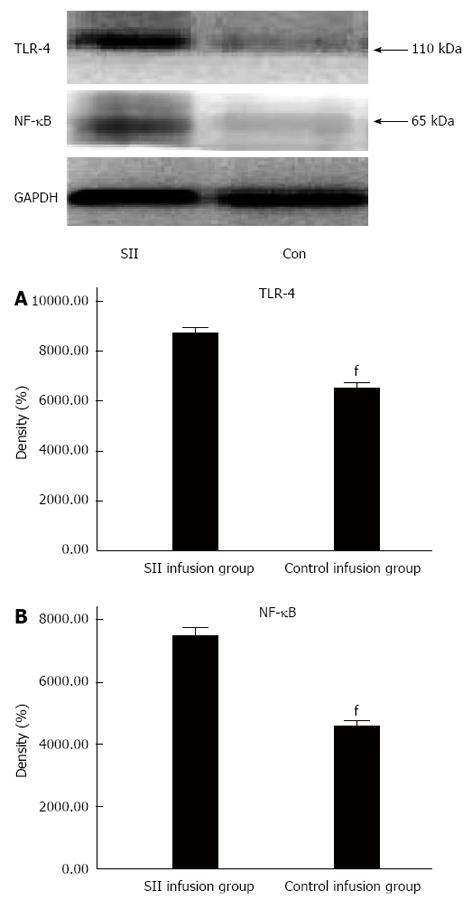
Figure 4 Compared with the control infusion group, toll-like receptor-4 and nuclear factor-κB expression levels were significantly increased in the severe intraperitoneal infusion group.
Density of Toll-like receptor (TLR)-4 (A) and nuclear factor (NF)-κB (B) in lung protein extracts of SII and control rats was examined using Western blotting. All results (mean ± SD) are representative of two experiments (n = 10 rats in each group). fP < 0.01 vs SII infusion group.
- Citation: Zhang YM, Zhang SK, Cui NQ. Intravenous infusion of mesenteric lymph from severe intraperitoneal infection rats causes lung injury in healthy rats. World J Gastroenterol 2014; 20(16): 4771-4777
- URL: https://www.wjgnet.com/1007-9327/full/v20/i16/4771.htm
- DOI: https://dx.doi.org/10.3748/wjg.v20.i16.4771


