Copyright
©2014 Baishideng Publishing Group Co.
World J Gastroenterol. Mar 28, 2014; 20(12): 3059-3068
Published online Mar 28, 2014. doi: 10.3748/wjg.v20.i12.3059
Published online Mar 28, 2014. doi: 10.3748/wjg.v20.i12.3059
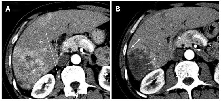
Figure 1 Application of modified Response Evaluation Criteria in Solid Tumor evaluation for hepatocellular carcinoma.
A: Baseline; B: Post treatment. Tumor burden change was assessed on arterial-phase contrast enhanced diagnostic computed tomography image. Modified Response Evaluation Criteria in Solid Tumor evaluation should draw the maximal dimension of continuous arterial enhancement in such lesions with central necrosis, avoiding central necrosis.
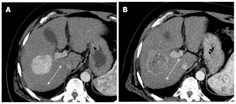
Figure 2 Portal-phase contrast enhanced diagnostic computed tomography of 58-year-old man with hepatocellular carcinoma.
A: Baseline; B: Post treatment. Obvious tumor density change was observed after antiangiogenic treatment.
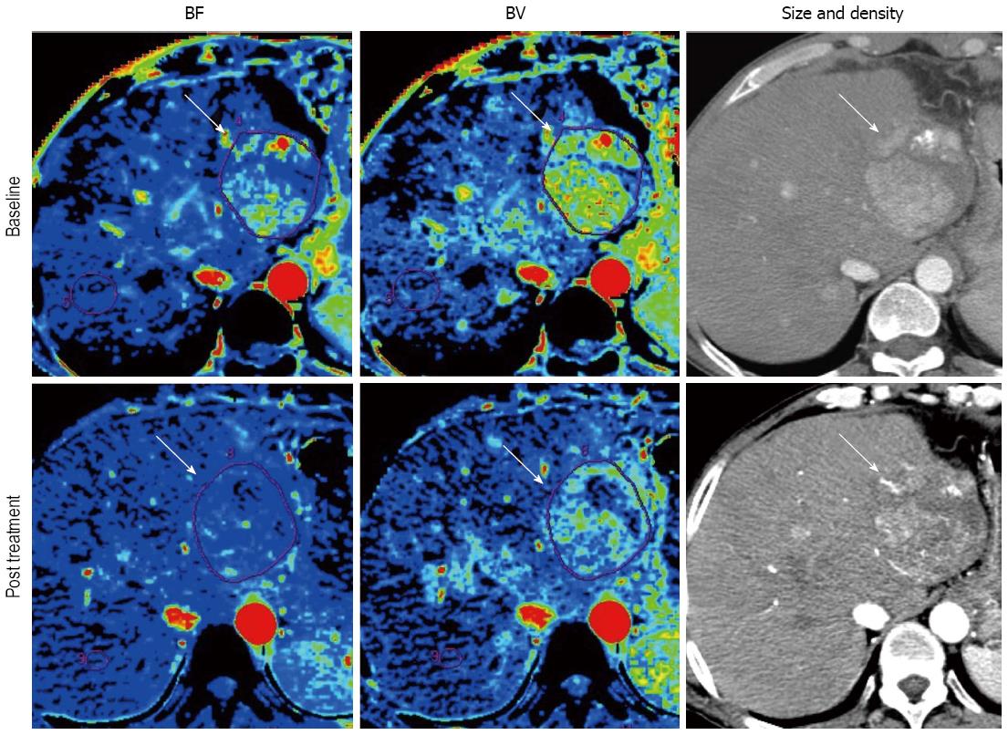
Figure 3 Perfusion maps of 53-year-old man with hepatocellular carcinoma.
Parameters measure by perfusion computed tomography showed substantial changes in comparison with tumor size and density at only 2 wk after antiangiogenic treatment. Blood flow (BF), blood volume (BV) were -75.5% and -59.5%. On the other hand, those of size and density were not so obvious (-3.0% and -18.1%).
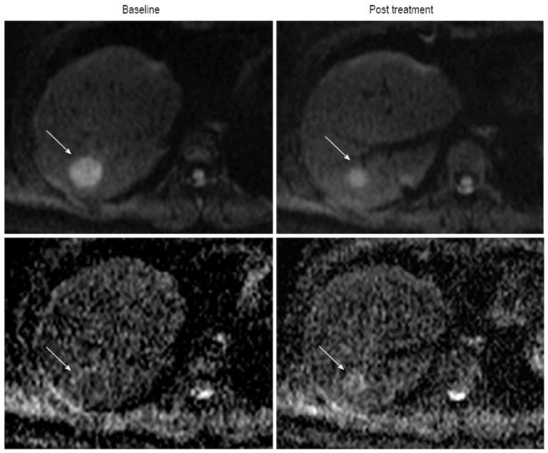
Figure 4 Diffusion-weighted imaging and apparent diffusion coefficient map at baseline and post treatment of 31-year-old woman with hepatocellular carcinoma (arrows).
This patient was treated with antiangiogenic agent (sunitinib). Apparent diffusion coefficient showed 18.8% increase (from 1.28 × 10⁻³ to 1.52 × 10⁻³ mm2/s) after antiangiogenic treatment.
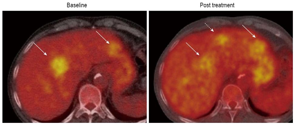
Figure 5 Positron emission tomography/computed tomography of 57-year-old man with hepatocellular carcinoma at baseline and post treatment.
He was treated with a systemic chemotherapy. New lesion was detected by a follow-up positron emission tomography/computed tomography (dotted arrow).
- Citation: Hayano K, Fuentes-Orrego JM, Sahani DV. New approaches for precise response evaluation in hepatocellular carcinoma. World J Gastroenterol 2014; 20(12): 3059-3068
- URL: https://www.wjgnet.com/1007-9327/full/v20/i12/3059.htm
- DOI: https://dx.doi.org/10.3748/wjg.v20.i12.3059


