Copyright
©2014 Baishideng Publishing Group Co.
World J Gastroenterol. Mar 21, 2014; 20(11): 3050-3055
Published online Mar 21, 2014. doi: 10.3748/wjg.v20.i11.3050
Published online Mar 21, 2014. doi: 10.3748/wjg.v20.i11.3050
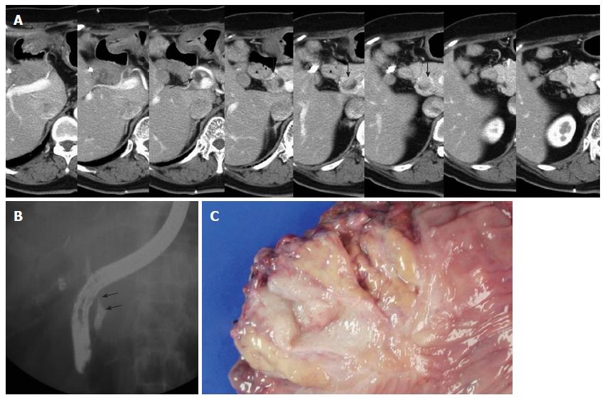
Figure 1 Imaging findings and gross specimen of the later metachronous bile duct tumor in case 1.
A: The arrow indicates the dilated remnant distal bile duct, and the arrow heads indicate the polypoid mass lesion of the distal remnant bile duct; B: Endoscopic retrograde cholangiopancreatography revealed mild dilatation of the intrapancreatic remnant bile duct with a filling defect (arrows); C: A gross examination of the resected specimen shows a polypoid mass arising from the remnant bile duct with invasion into the pancreas.
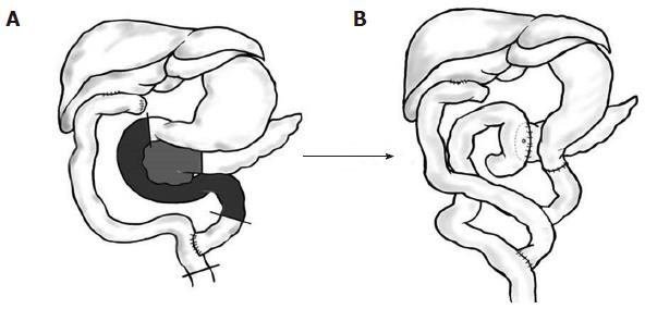
Figure 2 Panel A shows the previous bile duct resection with a Roux-en-Y hepaticojejunostomy.
The lines in panel A denote where the cut was made, and the shaded area indicates the part to be resected. Panel B shows the later pyloric preserving pancreatoduodenectomy with a Roux-en-Y hepaticojejunostomy.
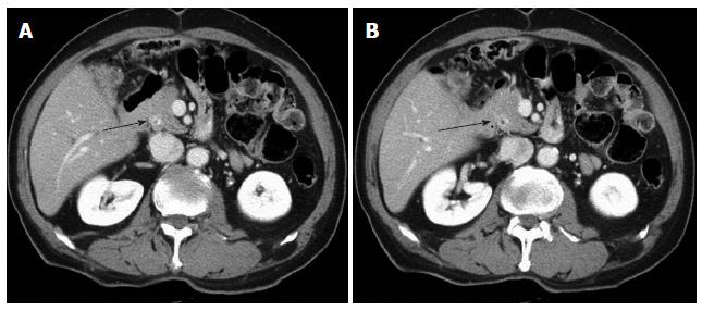
Figure 3 Abdominal computed tomography findings of the later metachronous bile duct tumor in case 2 (A, B).
The arrows indicate the new lesion with slight contrast enhancement at the distal remnant bile duct, but no definite mass was observed.
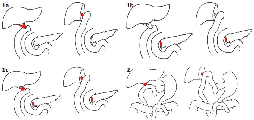
Figure 4 Classification of metachronous bile duct carcinoma according to the site of de novo neoplasia.
Type 1 tumors occur at the proximal and/or distal remnant bile ducts. Type 1a tumors with involvement of the proximal remnant bile duct only require hepatectomy. Type 1b tumors with involvement of the distal remnant bile duct only require pancreaticoduodenectomy. Type 1c tumors with involvement of both proximal and distal remnant bile ducts require hepatectomy and pancreaticoduodenectomy. Type 2 tumors with involvement of the proximal remnant bile duct after pancreatoduodenectomy require hepatectomy.
-
Citation: Kwon HJ, Kim SG, Chun JM, Hwang YJ. Classifying extrahepatic bile duct metachronous carcinoma by
de novo neoplasia site. World J Gastroenterol 2014; 20(11): 3050-3055 - URL: https://www.wjgnet.com/1007-9327/full/v20/i11/3050.htm
- DOI: https://dx.doi.org/10.3748/wjg.v20.i11.3050


