Copyright
©2014 Baishideng Publishing Group Co.
World J Gastroenterol. Mar 21, 2014; 20(11): 2751-2759
Published online Mar 21, 2014. doi: 10.3748/wjg.v20.i11.2751
Published online Mar 21, 2014. doi: 10.3748/wjg.v20.i11.2751
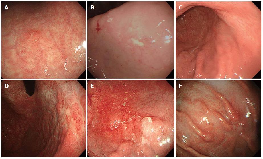
Figure 1 Variable endoscopic findings of gastric mucosa-associated lymphoid tissue lymphoma.
A: Single erosive type; B: Ulcerative type; C: Atrophic type; D: Cobblestone-mucosa type; E: Nodular type; F: IIc-like type.
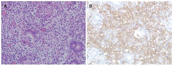
Figure 2 Histopathological and immunohistochemical features of gastric mucosa-associated lymphoid tissue lymphoma.
A: Histopathological finding. Gastric MALT lymphoma shows many lymphoepithelial lesions formed by the invasion of glands by centrocyte-like cells [hematoxylin and eosin (HE) stain, × 400]; B: Immunohistochemical finding. Centrocyte-like cells show strong positive signals for CD20 (× 400).
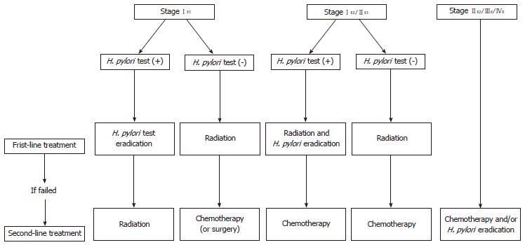
Figure 3 Ann Arbor staging system modified by Musshoff and suggested therapeutic algorithm for gastric mucosa-associated lymphoid tissue lymphoma.
H.pylori: Helicobacter pylori.
-
Citation: Park JB, Koo JS.
Helicobacter pylori infection in gastric mucosa-associated lymphoid tissue lymphoma. World J Gastroenterol 2014; 20(11): 2751-2759 - URL: https://www.wjgnet.com/1007-9327/full/v20/i11/2751.htm
- DOI: https://dx.doi.org/10.3748/wjg.v20.i11.2751


